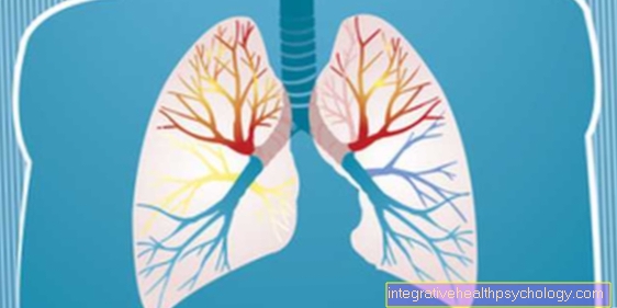
Atelectasis
Synonyms
Ventilation deficit, collapsed section of the lung
introduction
As "Atelectatic" is a part of the lung that is not ventilated. This part contains little or no air in its alveoli. A segment, lobe, or even an entire lung can be affected.
In order to function properly, the lungs must be well supplied with blood and well ventilated. This is the only way to ensure an exchange of substances between blood and air, in which the body can exhale sufficient CO2 and absorb sufficient oxygen.
If a part of the lungs has collapsed and is no longer filled with air, it can no longer contribute to breathing.
However, to understand how this can happen, it is first important to understand how breathing normally works.
causes
A distinction is made between innate (fetal, primary) Atelectasis and those acquired as a result of an unfavorable circumstance (secondary).
Congenital atelectasis can arise due to central nervous malfunctions, malformations or, in premature infants, a surfactant deficiency. Surfactant is a mixture of water, fats and proteins that is produced by the lungs in order to reduce the surface tension of the fluid layer in the alveoli to such an extent that it can develop in the first place. The production of this mixture begins quite late in the lung maturation.
Acquired atelectasis can have many causes.
In the so-called compression atelectasis, the collapsed area of the lungs is pressed by something and thus prevented from unfolding. This pressure can be exerted, for example, by a swelling (tumor), an accumulation of blood, pus or water in the gap between the lungs and the chest wall (pleural gap) or swollen lymph nodes. An injury to the chest wall or the lungs, in which air enters the gap between the lungs and the chest wall, can also compress the lungs. This form of atelectasis affects an entire lung, is also called relaxation atelectasis or pneumothorax, and is a serious condition.
In contraction atelectasis, the lack of ventilation is due to scarring of the lungs at this point, which in turn is the result of a lung disease such as tuberculosis or sarcoid.
In the case of microelectasis, for example in a shock situation, the lung tissue in the affected area was too poorly supplied with blood so that it could not produce sufficient surfactant. The surface tension of the fluid in the alveolar sacs (alveolar fluid) then pulls the lungs together in place.
Resorption atelectasis occurs when the air in one section of the lung is completely absorbed into the blood. This is conceivable if a patient is ventilated with pure oxygen for more than 3 minutes and then there is almost only oxygen in the alveoli. Obstruction atelectasis is a subtype of resorption atelectasis. This occurs when a branch of the lung (bronchus) is pinched off and the air trapped behind it is absorbed into the blood over time. In turn, such clamping may be caused by a tumor, swallowing an object, or swollen lymph nodes.
Read more on the topic:
- Lung disease
- Respiratory distress syndrome in premature infants
Symptoms and consequences
Depending on how an atelectasis develops and how large the affected lung area is, the development and re-resolution of an atelectasis can either go unnoticed or be associated with pain, cough and severe shortness of breath. The development of a so-called pneumothorax is often painful.
Since there is a lack of oxygen in the affected areas, the blood circulation is also reactively throttled by constricting the vessels (Euler-Liljestrand mechanism). This is to ensure that blood flowing through the lungs is really rich in oxygen afterwards. However, this constriction (vasoconstriction) also increases the resistance against which the right heart has to pump, which can lead to further problems, especially in the case of pronounced atelectasis or pre-existing cardiac insufficiency. Furthermore, the parts of the lungs with poor blood circulation are more susceptible to infections and inflammations, e.g. Pneumonia, including water deposits (edema), are more likely.
If the blood in the lungs is no longer loaded with enough oxygen, so-called cyanosis with blue discoloration of the fingernails, lips and tongue can occur.
The clinical picture of tension pneumothorax, in which the accumulation of air around the lungs through a valve mechanism increases with each breath, is life-threatening.
diagnosis
During the medical examination, atelectasis presents with a dampening of the knocking sound, when listening (Auscultation) a weaker breathing sound is noticed. With the help of an X-ray image, a CT or ultrasound examination, atelectasis is primarily characterized by the decrease in volume of the affected area and an associated increase in density. In the case of a larger atelectasis, the surrounding structures can shift towards this.
Recently, the MRI of the lungs with helium has been added as a newer diagnostic method.
What does an atelectasis look like in an X-ray?
Atelectasis appears in the X-ray image as uniform shading, which is based on the boundaries of the lung lobes.
The radiological findings show a reduction in lung volume due to the evacuated areas of the atelectasis. Depending on the extent of the atelectasis, the diaphragm is also elevated and the lungs and trachea shift to the affected side.
therapy
Small atelectases usually go away on their own or with the help of repositioning and breathing exercises, and intervention is especially necessary for larger events. In the case of compression atelectasis, the constricting element (air, blood, pus, water) is removed with the aid of a draining tube. Ventilation with positive pressure is also conceivable in order to bring the compressed lung sections to unfold again. Oxygen can be added to the breathed air symptomatically.
In the case of atelectasis, however, it is particularly important to always clarify the cause, as atelectasis can be a sign of a serious illness.
Read more on this topic: Chest drain
Plate atelectasis
The so-called plate atelectases are flat, a few centimeters long, strip-shaped atelectases that are not bound to the lung segments and are often located above the diaphragm in the lower lung segments. Plate atelectasis occurs particularly in diseases of the abdominal cavity, for example as a result of an abdominal operation with subsequent bed rest and insufficient breathing or ventilation of the lungs.
But they can also occur in connection with pneumonia, a heart attack, whooping cough or as a result of a malformation of the chest.
What is atelectasis prophylaxis?
Patients who have recently had surgery, are immune to weak immune systems and suffer from respiratory diseases, as well as elderly, debilitated and especially bedridden patients, are at risk of developing atelectasis in certain parts of the lungs.
To prevent this, breathing exercises should be carried out regularly. Since patients in the above-mentioned In situations or patients with chronic lung disease often because of the circumstances they have improper breathing technique or inefficient breathing, respiratory physiotherapy teaches certain techniques to improve breathing.
By strengthening the respiratory muscles and improving the efficiency of breathing, sections of the lungs are ventilated that would otherwise be less ventilated and are at risk of developing atelectasis.
In addition to breathing exercises that are carried out regularly, the patient's mobilization, sufficient fluid intake and regular repositioning play a major role in the prevention of atelectasis.
forecast
The chances of recovery from atelectasis are usually very good; secondary manifestations are in principle always reversible. Pronounced forms, such as tension pneumothorax, can be treated very well, but if left untreated, they can lead to death.
Respiratory physiology
In the healthy lungs, fresh air is brought together with blood from the body at the smallest level, only separated by the unimaginably thin wall of an alveoli, in which the air is located, and the likewise wafer-thin wall of the fine veins (capillaries) in which the blood is contained the air bubble flows around. The concentrations of CO2 and oxygen in blood and air can now be brought into line via this thin barrier. The CO2-rich blood from the body releases this into the CO2-poor air; in return, oxygen (O2) from the air enters the blood, which has previously released its oxygen to the body. The concentration difference is maintained through constant breathing and the flow of blood and a continuous gas exchange is possible.
The lungs themselves, through elastic components in the lung tissue as well as through the surface tension of the layer of fluid lining the alveoli, constantly strive to contract, i.e. to "collapse". It is prevented from doing this by the fact that there is a negative pressure between the lungs and chest wall, which always pulls them apart. When inhaling, the lungs are expanded further by lowering the diaphragm and expanding the chest.