Task of the heart
introduction
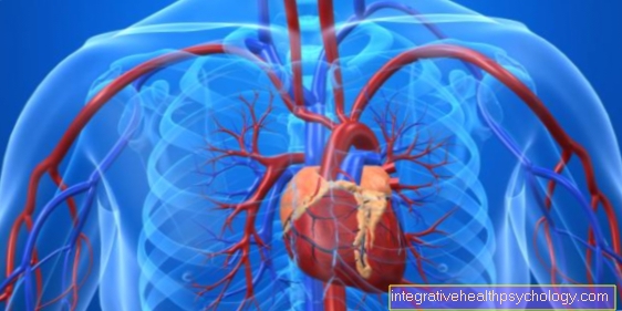
The heart plays a central role in the human cardiovascular system because it is the motor of the circulatory system.
The blood from the body's circulation first reaches the right half of the heart. From there, the blood is pumped to the lungs, where it is supplied with oxygen. The blood flows from the pulmonary circulation into the left half of the heart, from where it is transported through the main artery into the entire body circulation. In its pumping function, the heart adapts to the respective situation of the body, because depending on whether a person is lying down, standing or exerting himself physically, different demands are made on the cardiovascular system.
Read more on the topic: Function of the heart
Functions of the left ventricle
The left ventricle, also called the left ventricle is responsible for pumping the blood into the body's circulation. In the Relaxation phase, the so-called diastole becomes the left ventricle with oxygenated blood from the pulmonary circulation filled. This first reaches the left atrium from the lungs. From there it is transported through the mitral valve into the left ventricle.
During the Tension phase, in medical terminology as Systole called, the left ventricle pumps the blood through the Aortic valve in the main arteryfrom where it enters the body's circulation.
The left ventricle must be like this apply a lot of pressurethat it exceeds the blood pressure in the body's circulation. Normally this is around 120 mmHg. Compared to the right ventricle, the left ventricle therefore has to apply a significantly higher pressure. That's why the Heart muscle layer on the left Side very much thicker.
The left ventricle must be able to adapt its pumping activity to the physical activity. During one physical exertion therefore the heart beats clearly more quickly. At the same time, the left ventricle is filled with a larger amount of blood before each beat. This increases by increasing the volume and the beat frequency Cardiac output, that is the amount of blood that the heart pumps into the circulation per minute. Only in this way is the heart able to meet the increased demands in a stress phase.
Functions of the right ventricle
The right ventricle gets the oxygen-poor blood from the body's circulation. This is first collected in the right atrium before it is transported through the tricuspid valve into the right ventricle. The right ventricle pumps the blood through the so-called pulmonary valve into the pulmonary vesselsso that it is supplied with oxygen in the lungs.
There are two different phases in the pumping function of the right ventricle. The Tension phase and the Relaxation phase.
During the Relaxation phase fills the right ventricle with blood, the heart muscles are relaxed. When the signal for tension arrives in the right ventricle via the cardiac conduction system, the heart muscle tenses. As soon as a pressure of about 25 mmHg is reached, this is the maximum pressure that prevails in the pulmonary circulation, the tricuspid valve opens. Thus, the blood comes out of the right ventricle in the pulmonary circulation.
In contrast to the left ventricle, the right ventricle has to apply a comparatively low pressure in the tension phase. This is why the muscle layer in the right ventricle is significantly thinner.
Functions of the auricles
In the Atria the heart collects the blood from the previous sections of the circulatory system. Through the upper and lower vena cava the blood from the body's circulation enters the right atrium.
From there it gets through the Tricuspid valve through into the right ventricle pumped. The forecourt itself hardly has its own pumping function. Rather, the blood gets through the negative pressure, which is created in the right ventricle during the relaxation phase, is sucked into it.
The blood that im left atrium lands, comes from the pulmonary circulation. From the left atrium it becomes through the Mitral valve pumped into the left ventricle.
Like the ventricles, the atria have one Tension and a relaxation phase. However, these run opposite to the phases of the ventricles. In the relaxation phase of the heart chambers, the atria must contract so that they can pump the blood into the chambers. While the chambers tense, the atria are already filling up again with the blood from the previous sections of the circulatory system.
Role of the heart in the circulatory system
The heart forms the Motor of the cardiovascular system. About 5 liters of blood pass through the heart every minute. That corresponds to the whole Blood volume.
In terms of blood flow, the heart is divided into a right and a left half. Colloquially one speaks of the "right" and "left heart". While the right half of the heart the blood from the entire body circulation collects and in the Pulmonary vessels pumps, comes in the blood from the pulmonary circulation on the left side of the heart and get back from there into the rest of the body.
Although both halves of the heart have to handle the same amount of blood, this is the left ventricle clear more muscular. This is because she has to keep pumping the blood against a higher pressure.
Depending on the state of the body, the heart must different requirements satisfy. In a person lying down, the heart has comparatively little to do. While standing, part of the blood has to be pumped into the brain against gravity. A little more strength is needed for this.
who Sports makes, drives Heart to top performance. Because during sport, the muscles of the body must also more nutrients and oxygen are supplied. To do this, the cardiovascular system needs to be stimulated, which means more work for the heart.
Task of the conduction system of the heart
So that the heart pumps blood reliably and evenly into the circulation, everyone must Coordinates muscle cells of the heart become. That's what it's for Excitation conduction system. This consists of annoy, the information only the next from one heart muscle cell transport.
The conduction system begins on Sinus node in the Atria. If the electrical signal reaches the muscle cells there, they tighten and continue to pump the blood into the heart chambers. The muscle cells then relax again. Meanwhile, the signal continues to run in the excitation conduction system. It passes through the heart septum to the tip of the two heart chambers and then, along the outer heart wall, back towards the heart base.
This also works in the ventricles Signal for the tension of the heart muscle cellswhich removes the blood both chambers in the circuit is pumped. While the electrical signal goes back in the heart chambers and the muscles relax there again, the signal for the next heartbeat is generated at the sinus node.
Task of the sinus node
Of the Sinus node is the pacemaker of the heart. That means that here the electrical impulses arisethat make the heart beat.
The sinus node sits in the right atrium. From there the excitement spreads to the AV node out and will then into the ventricles forwarded. Usually the sinus node gives one rhythm from about 60 to 80 beats per minute.
At burden for example through physical exertion, in stressful situations or in fear, the heart beats faster. In order to control these processes, the sinus node receives information from the brain and converts it in the form of faster or slower impulses.
AV node
Of the AV node has a guardian function in the conduction system of the heart. The excitation of the heart muscle cells spreads from the sinus node over the atria and ends up at the AV node. This directs the given Heart rate into the ventricles further.
A important function come to that AV node toowhen the sinus node is no longer working properly. If the impulse is too fast, which is the case with atrial fibrillation, for example, the AV node does not pass all the excitations on to the ventricles. Consequently he controls the frequency and ensures that the chambers continue to operate at a normal speed. The sinus node falls completely out, jumps of the AV node. He then generates the impulses that make the heart beat. However, the heartbeat is a little slower in this case.
Task of the heart valves
The heart Has four heart valves, where one Pocket and sail flaps differs.
The two leaflet valves separate the atria of the heart from the ventricles. The so-called Tricuspid valve lies between the right atrium and the right ventricle, which Mitral valve forms the border between the left atrium and the left ventricle. These two heart valves are in the Tension phase of the heart closed and thereby prevent the blood from being pumped backwards in the heart. In the During the relaxation phase, the two wing flaps openso that the heart chambers can fill up with blood again.
The pocket flaps separate the heart chambers from the vascular system behind. The Pulmonary valve lies between the right ventricle and the pulmonary circulation. The Aortic valve separates the left ventricle from the body's circulation. Both Valves are in the relaxation phase, also the filling phase, as the heart chambers fill up, closed. When the heart is tense, these heart valves are opened and the blood enters the circulation.
The flaps become problematic when they are leaking. This is then called Heart valve insufficiency designated. In addition, there may be a narrowing in the area of the valves, in technical language this is Stenosis called. In both cases, the heart has to do more work.
Task of the coronary vessels
The human heart is about two main vessels supplied: the left and the right Coronary artery. The coronary arteries or in technical terms Coronary arteries, are responsible for the To supply blood to the heart muscle.
This will make enough Transports nutrients and oxygen to muscle cells. In addition, those arising from the work of the heart Waste materials from the muscles again transported away.
In most muscles, the supplying vessels form a large number of cross-connections. As a result, the entire muscle continues to be supplied with sufficient blood even in the event of a blockage in a vessel. The coronary vessels also form these so-called Collateral circuits out. However, these are not enough to fully supply the heart. That's why the Closure of a branch of the coronary arteries to a Undersupply the muscles behind it.
Due to the pumping activity of the heart, the coronary vessels are not supplied with blood all the time. Only in the Relaxation phase of the heart blood gets into the vessels. The heartbeat becomes faster during exercise, which also means that the relaxation phase is shortened. As a result, the duration in which the coronary arteries are supplied with blood is also shorter.
Task of the heart septum
The Cardiac septum is the part of the heart that zwipe the two chambers of the heart lies.
So your first task is to meet the two of you Separate chambers from each other. Like the outer parts of the heart, the heart septum is made of Musculature and, together with the rest of the heart, pumps the blood into the circulation during the tension phase. Since the left ventricle has to do significantly more work than the right, the heart septum mainly supports the left ventricle in their pumping function. Also run in the heart septum Parts of the excitation conduction system. In it, the electrical signal runs from the atria to the tip of the heart.
Task of the pacemaker
A Pacemaker is needed when the heart is there can no longer do it alone, regularly to beat. This can have various causes. For example, the Sinus node, the heart's own pacemaker, no longer reliable or there is Problems in the conduction system. In both cases the pacemaker can do the take on the missing function.
There are different Types of pacemakers. Those who replace the function of the sinus node are responsible for one to establish an electrical signalwhich is then sent through the entire conduction system of the heart.
Other pacemakers provide the Connection between the atria and the ventricles again. In this case, a signal that arrives from the atria is passed on via the AV node to the ventricles. This allows the heart chambers to continue working in the usual way. The latest pacemaker can also Cardiac activity disorders, so-called cardiac arrhythmias, directly record. In addition, today's pacemakers are able to adapt the heart rate to the respective physical activity of the person.
Task of the pericardium
Of the Pericardium almost completely encloses the heart. Only the vessels that come directly from the heart (pulmonary artery and main artery) go through it.
The most important task of the pericardium it is the heart to protect. In addition, this is connected to the surrounding tissue and thus gives the heart its support in the chest. The pericardium consists of two different layersbetween which there is about 10-15 ml of liquid. As a result, the two layers can be moved relative to one another and allow the heart to move freely. This is necessary so that the heart can contract and relax. However, there is not unlimited space in the pericardium. If to much liquid or one some amount of blood between the pericardium and heart the heart is clearly restricted in its pumping function, as it can no longer expand completely.