Knee joint
- Anatomy knee joint
- Figure cruciate ligaments
- X-ray knee joint
- Osteoarthritis of the knee
- Meniscus and meniscal tear
- Cruciate ligament injury
- Collateral ligament tear / inner ligament stretch
Synonyms
Articulatio genus, knee, thigh roll, tibial head, joint, femur, tibia, fibula, kneecap, meniscus, cruciate ligaments, anterior cruciate ligament, posterior cruciate ligament, collateral ligaments, inner ligament, outer ligament
Figure knee joint
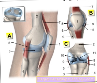
A - Right knee joint from the left
B - Right knee joint from the front
C - Right knee joint from behind
- Kneecap - patella
- Femur - Femur
- Shin - Tibia
- Fibula - Fibula
- Inner meniscus -
Meniscus medialis - Outer meniscus -
Lateral meniscus - Kneecap ligament -
Ligamentum patellae - Outer band -
Ligament collaterale fibulare - Inner band -
Ligament collateral tibial - Posterior cruciate ligament -
Ligament cruciatum posterius - Anterior cruciate ligament -
Ligament cruciatum anterius
You can find an overview of all Dr-Gumpert images at: medical illustrations
Anatomy knee joint
- Thigh muscles (Musculsus quadriceps femoris)
- Thighbones (Femur)
- Thigh tendon (Quadriceps tendon)
- Kneecap (patella)
- Kneecap tendon (Patellar tendon)
- Kneecap tendon attachment (Tibial tuberosity)
- Shin (Tibia)
- Fibula (Fibula)
anatomy
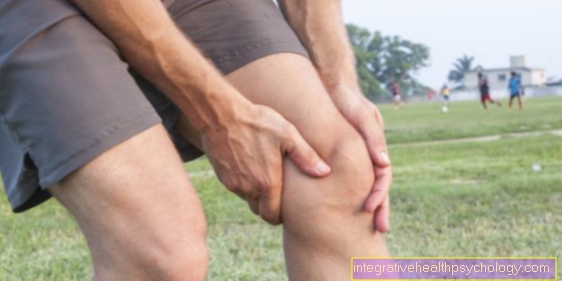
The Knee joint is the largest and most stressed joint of the human body. Accordingly, the knee is also the most commonly injured joint.
At the Knee joint it is a so-called rotary hinge joint. This means that the knee joint can be both bent and rotated
The knee joint becomes three bone formed, the Thigh bone (femur), the Shinbone (tibia) and the Kneecap (patella). The Fibula is not involved in the knee joint.
The knee joint is differentiated into two lower joints:
- the Femoral and shin joint (Femorotibial joint)
- the Shin - kneecap - joint (Femoropatellar joint)
The Thigh - shin - joint
The joint is formed by the thigh bone with its two joint heads (medial and lateral femoral condyle) and the tibial plateau (tibial plateau). The round heads of the thighs lie in the small hollows of the shinbone plateau (intercondylar fossa). The ratio of the joint surface of the thigh to the shin is about 3: 1.
Since there is only a point-like contact between the thigh and the shin, the diffraction of the knee joint to a rolling sliding movement.
The Thigh - kneecap - joint
When you bend, the kneecap slides through a predetermined slideway between the thighbone heads (femoral condyles). Overall, the kneecap can slide between 5 - 10 cm. In order to cover this distance, larger sliding layers are necessary. For this purpose, two bursa (prepatellar bursa and infrapatellar bursa) form two large sliding gaps. The large thigh muscles (Musculus quadrices femoris) attach to the kneecap (patella) from above. The strength of these muscles is about the Kneecap (Patella) diverted to the lower leg. The kneecap tendon (patellar tendon) attaches to the lower kneecap pole, pulling towards the front edge of the tibia and connecting to the tibia at a protruding bone (apophysis = tibial tuberosity).
Various knee stabilizers are available to stabilize the thigh in the small socket of the tibia (intercondylar fossa):
- the meniscus (inner meniscus and outer meniscus)
- the cruciate ligaments (anterior cruciate ligament, posterior cruciate ligament)
- the side ligaments (inner ligament, Outer band)
- the joint capsule
The menisci (plural of the meniscus) help with the transfer of force from the thigh to the lower leg. Since the head of the femur is round and the tibia plateau is almost straight, there is only point contact. To increase the contact area, there is the Medial meniscus and the External meniscus. They are inserted on the inside and outside as a kind of shock absorber and help to distribute the force more evenly. You can find more about this in our articles Meniscus and Meniscus lesion (Meniscus damage).
The cruciate ligaments prevent the femoral heads from sliding forward relative to the shin (anterior cruciate ligament) or to the rear (posterior cruciate ligament). They are the decisive stabilizers of the knee joint.
The collateral ligaments stabilize in a lateral direction so that the knee joint cannot buckle into a bow-leg or knock-kneel malposition. The inner ligament is firmly fused with the inner meniscus, so the inner meniscus is more immobile than the outer meniscus.
The joint capsule of the knee joint is very tense and stabilized when it is fully extended. With increasing flexion it slackens and the remaining stabilizers have to take over the tasks.
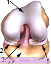
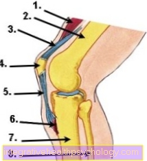
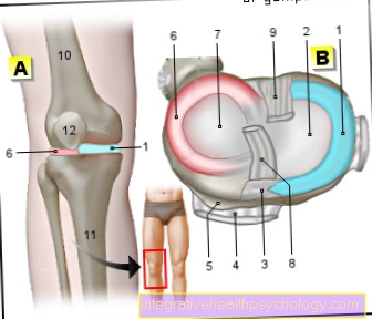
Figure cruciate ligaments
- Thighbones (Femur)
- Medial meniscus
- anterior cruciate ligament (VKB)
- Shin (Tibia)
- External meniscus
function
Normally the knee can be bent up to 120 - 150 ° and, depending on the ligamentous apparatus, about 5 - 10 ° overstretched. With 90 ° flexion, the knee can be rotated approximately 40 ° outwards and 10-20 ° inwards.
The knee joint must bear the entire load of the trunk on the lower leg (Shin = Tibia).
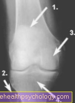
X-ray knee joint
taken from the front (a.p.)
- Kneecap (patella)
- Fibula (Fibula)
- Thighbones (Femur)
- Shin (Tibia)
Diseases

No joint in the human body has more injuries than the knee joint.
Osteoarthritis of the knee
The most common disease of the knee joint is Osteoarthritis of the knee. This results in damage to the cartilage sliding layer of the knee joint. Can follow inflammation, Pain, restricted mobility and instability.
Further information is available under our topic:
- Osteoarthritis of the knee
- Cartilage damage in the knee
Meniscus and meniscal tear
In addition to the cartilage damage, this occurs especially with increasing age Tears in the meniscus more often. Due to the lack of healing potential of the meniscus, there is only a partial removal of the torn meniscus, apart from a few exceptional cases.
Further information is available under our topics:
- Meniscal tear
- meniscus
Cruciate ligament injury
One is rarer, but usually more serious Cruciate ligament injury, as you significantly reduce the stability of the knee joint. In comparison, the anterior cruciate ligament injury is more common than the posterior one.
Further information is available under our topic:
- Cruciate ligament injury
Collateral ligament tear / inner ligament stretch
Torn collateral ligaments often occur in combination with other injuries. While inner ligament damage has good healing potential, it must be complete Outer ligament tears are usually treated surgically.
Further information on the subject can be found at: Inner ligament stretch in the knee
Pain in the knee joint
Knee joint pain can have various causes.
Depending on the localization of the pain, this can occur different diseases or injuries Clues.
Also the point in time at which the pain preferentially occurs (at rest, at night, as initial pain, during exertion), can provide further clues as to the underlying cause.
Internal knee pain:
Knee pain, mostly on the inside of the knee, is often indicative of one Meniscus damage down.
Especially the one Medial meniscus is often injured in many sports injuries. This can be noticed by pain on the inside of the knee.
Also one Leg malposition (If one) can permanently damage the inner meniscus, as the pressure on it is increased by the uneven load. Ultimately, this can result in a Osteoarthritis in the knee joint train, which can express itself through internal knee pain.
Ultimately, a Bursitis or a muscular damage that cause discomfort.
External knee pain:
Pain on the outside of the knee can also be caused by a Leg malposition (X-leg) be conditional. In this case, the external meniscus is stressed more than normal.
This can permanently damage the meniscus and subsequently Osteoarthritis development to lead. This can be responsible for the pain.
Another cause of outside knee pain is one Damage to the hamstring tendon (Biceps femoris).
Knee pain on the outside of the knee in runners can also be attributed to the so-called Runner's knees indicate a pain syndrome caused by the Overuse of the iliotibial band, a fascia band on the outside of the thigh.
The symptoms typically only occur when running, and later when walking.
Front knee pain:
Pain that is mainly felt in the front area of the knee can be attributed to a Damage to the patella tendon Clues.
Also one Malformation of the kneecap (Patellar dysplasia) can cause these complaints, as in this case the kneecap does not fit optimally into its abutment and can cause pain due to friction and incorrect loading.
A Bursitis of the knee can also cause pain in the anterior knee area.
Back knee pain:
Pain in the hollow of the knee can have multiple causes. These include vascular, bone and nerve damage, ligament or meniscus injuries, as well as one Baker's cyst.
This is a protuberance of the joint capsule of the knee joint, which is increased by Synovial fluid arises. The production of synovial fluid is often significantly increased by chronic inflammatory processes, for example in the context of a rheumatic disease.
When a Baker's cyst rips, it can become a dangerous one Compartment syndrome come.
Generalized knee pain:
If the pain is diffuse throughout the knee joint, it may be one inflammation, one rheumatic or one degenerative disease (arthrosis) act.
The knee joint is common in parallel to an inflammatory process swollen, overheated and if necessary reddened.
Especially Starting pain and Stiffness after a longer period of rest, this can speak for knee osteoarthritis.
For systemic signs of inflammation, such as fever or poor general condition it is more likely that it is an acute inflammatory process.
In any case, a visit to a doctor is advisable if the symptoms persist for a long time.

I would be happy to advise you!
Who am I?
My name is dr. Nicolas Gumpert. I am a specialist in orthopedics and the founder of .
Various television programs and print media report regularly about my work. On HR television you can see me every 6 weeks live on "Hallo Hessen".
But now enough is indicated ;-)
The knee joint is one of the joints with the greatest stress.
Therefore, the treatment of the knee joint (e.g. meniscus tear, cartilage damage, cruciate ligament damage, runner's knee, etc.) requires a lot of experience.
I treat a wide variety of knee diseases in a conservative way.
The aim of any treatment is treatment without surgery.
Which therapy achieves the best results in the long term can only be determined after looking at all of the information (Examination, X-ray, ultrasound, MRI, etc.) be assessed.
You can find me in:
- Lumedis - your orthopedic surgeon
Kaiserstrasse 14
60311 Frankfurt am Main
Directly to the online appointment arrangement
Unfortunately, it is currently only possible to make an appointment with private health insurers. I hope for your understanding!
Further information about myself can be found at Dr. Nicolas Gumpert
Tap the knee joint
To stabilize the knee joint, it can be helpful to put tape around it. This method is particularly useful for follow-up treatment after knee injuries, as the tape movement supportive works, but does not restrict movement.
It also has one pain relieving effect and gently restores the knee to its normal functionality.
There are a few things to consider when taping the knee joint. First, the tapes tailored become. It will two shorter ones and two longer strips needed.
To measure the optimal length, use the Kneecap. The shorter tapes should end about three fingers wide above and below the kneecap, the two longer tapes about six fingers wide, i.e. three fingers wider than the shorter tapes.
Then the knee is reduced by about Bent 70 ° and the shorter tapes are glued tightly around the kneecap. It must full train be applied to the tapes, but do not pull the skin when sticking them, otherwise blisters may form; so always make sure that the ends of the tape are loosely attached to the skin.
The longer tapes glued. These will NOT under tension but loosely glued to the knee next to the shorter inner tape strips. The strips should overlap about a finger's breadth.
When the knee is then moved, the skin over the kneecap should wrinkle significantly in the extended position. The tape shouldn't feel uncomfortable. Overall, it is important to note that the inner strips are glued tight enough around the kneecap and the outer strips are applied around them completely without tension. This is the only way for the kinesiotape to develop its optimal stabilizing effect.
Read more on this topic at: Tap your knees
Knee joint surgery
An operation on the knee joint can be necessary in the event of various damage or diseases of the knee joint, if conservative therapeutic measures have not been successful.
Meniscus operations:
A meniscus injury can occur in the course of sports accidents.
If the meniscus is torn, it is often necessary to remove it to sew. However, this only works for not too large cracks and tears in a well-perfused area of the meniscus, as otherwise the healing process may not be adequate.
In such cases the torn meniscus part removed and be replaced by synthetic or natural material (Meniscus transplant).
Cruciate ligament surgery:
Cruciate ligament injuries also often have to be operated on.
A torn cruciate ligament leads to instability of the knee joint and can lead to the development of consequential damage and a Osteoarthritis of the knee to lead.
The standard procedure nowadays is autologous transplant an endogenous tendon to replace the injured cruciate ligament. The tendon of the Semitendinosus muscle used.
Is the cruciate ligament with a piece of bone torn off, this piece of bone including the cruciate ligament can be returned to its original position screwed become. This is particularly common in children and adolescents. An autologous transplant is then not necessary.
Same goes for one only torn cruciate ligament. This can often be reconstructed and sutured without the need for additional tendon tissue.
Cartilage transplant:
A novel treatment concept exists for cartilage damage in the knee joint. Be there the body's own cartilage cells are taken, bred and secondarily transplanted back into the knee jointwhere the cells can grow and compensate for cartilage defects.
Knee prosthesis:
Especially in the context of a serious Osteoarthritis of the knee In the long term, irreversible damage to the joint can occur, so that normal function is no longer possible.
If all conservative treatment measures have been exhausted, the last resort can be Knee endoprosthesis (Knee replacement) can be used. The knee joint is completely through artificial material replaced.
This is followed by a intensive physiotherapyso that the new knee joint is optimally resilient and the body can get used to it.
Lateral retinaculum split:
This procedure on the knee joint is performed when a Misalignment of the kneecap present.
In this case, the kneecap is pulled too much outward by the ligamentous apparatus and thus leads to increased pressure load on the outer part of the joint. This can cause consequential damage in the long term.
By a Splitting of part of the lateral ligamentous apparatus, the lateral retinaculum, the tension on the kneecap is reduced so that it is shifted more towards the center.
This distributes the force more evenly across the knee joint.
Kneecap surgery:
The so-called kneecap surgery according to Blauth is used when the lateral splitting of the retinaculum has not improved the symptoms.
The aim here is that the kneecap is shifted more towards the center and the pressure can be distributed more evenly on the joint.
To do this, the Patellar tendon severed and shifted further inwardsso that the kneecap is pulled towards the center.
Knee joint type of joint
The knee joint is a compound joint. On the one hand, it consists of the Kneecap joint (Femoropatellar joint) and on the other hand from the Popliteal joint (Femorotibial joint).
The popliteal joint is the actual knee joint, which enables the knee to be bent. Again, it's a combination of one Hinge- and one Wheel joint and is therefore also called Swivel joint designated.
Correspondingly executable movements are one Elongation and diffraction, as well as one in the bent state Outside- and Internal rotation of the knee.
The kneecap joint is also called Slide joint because the kneecap only slides in a bony groove at the lower end of the thigh bone. It is held in place by ligaments and slips over the cartilaginous joint surface when the knees are bent and extended.
Since the knee joint is exposed to great stress, it needs additional stabilizers.
Hence it is with the front and posterior cruciate ligament, as well as the Outside- and the Medial meniscus fitted. The menisci buffer shocks and ensure a more stable connection between the upper and lower legs.
Knee joint ligaments
So that the knee joint can withstand the daily loads, it is stabilized by numerous ligaments. Depending on their location, these are divided into a front, rear, side and central group. In sports injuries, the ligaments are not infrequently affected.
Front ligaments:
The front ligaments include that Kneecap ligament (Ligamentum patellae) and the Patellar retinaculum.
The patella ligament connects the kneecap to the front surface of the tibia. It is therefore very important for the power transmission from the upper to the lower leg during the stretching movement in the leg.
The patellar retinaculum lies to the side of the kneecap and secures it in position. It is also part of the with its various proportions Joint capsule of the knee joint.
Back ligaments:
At the back of the knee there are two other ligaments that stabilize the knee joint, on the one hand that sloping back of the knee (Ligamentum popliteum obliquum) and on the other hand that arched popliteal ligament (Arcuate poplite ligament). They are also part of the joint capsule.
Side straps:
The lateral ligaments of the knee joint are also called Collateral ligaments designated.
The inner collateral ligament runs inside the knee (Ligamentum collaterale tibiale), the outer collateral ligament runs on the outside of the knee (Ligamentum collaterale fibulare).
They stabilize the knee especially in the Stretching positionbecause they are relaxed when the knee is bent. This prevents the knee from slipping sideways when the knee is extended.
The bands work one O- or X-leg deformity opposite. It is important that that The inner ligament is firmly fused with the joint capsule and the inner meniscus while the outer ligament has no fixed connection to the joint capsule or the outer meniscus.
Therefore, an injury to the medial ligament is often accompanied by an injury to the medial meniscus. If the anterior cruciate ligament is also injured at the same time, one speaks of a "unhappy triad“.
Central bands:
The two cruciate ligaments (Ligamentum cruciatum anterius and posterius) educate the central tape backup of the knee joint. They cross over between the upper and lower leg bones.
Their position stabilizes the knee in the frontal plane, thus preventing the bones from sliding back and forth against each other. They also inhibit internal rotation (Inward rotation) of the leg.
Knee joint inflammation
Inflammation of the knee joint can have different causes. For example, it can be caused by an injury Wear processes (degeneration), by a Autoimmune disease or by a Infection with pathogens caused.
Ultimately it comes to one Inflammatory response in the knee joint, which is through a swelling, overheat, Redness and Pain expresses. As a result, the function of the knee joint is often significantly restricted.
Especially at bacterial infections of the knee can have symptoms like general feeling of illness and fever to be added.
To determine the exact cause of knee inflammation, the Joint effusion to be punctured. The fluid is withdrawn from the knee joint with a cannula and can then be examined for possible pathogens.
The problem with inflammation of the knee joint is that it can cause permanent damage to the joint. The cartilage in particular is attacked by the inflammatory reaction.
Has the damage progressed so far that Painkiller and anti-inflammatory drugs can no longer provide relief and other joint-preserving measures do not bring any improvement, the replacement of the knee joint with a prosthesis should be considered.
For this reason, knee pain that persists over a longer period of time should always be clarified by a doctor. In this way, suitable therapy can be initiated at an early stage and long-term damage avoided.
Knee joint bursa
Bursa serve the Cushioning of mechanical stress, as well as improving the gliding ability of tendons and ligaments.
In the knee area there are several bursa because the knee is heavily used every day and this way it can be relieved. A large bursa (Prepatellar bursa) is located between the kneecap and the overlying skin.
She serves the The ability of the skin to slide on the kneecap when flexed of the knee. The suprapatellar bursa is also known as the suprapatellar recess. It is another bursa and lies between the lower end of the thigh bone and the Quadriceps tendon.
This will make that smooth gliding of the tendon over the bone when flexed of the knee. Finally, the infrapatellar bursa lies beneath the patellar tendon and allows it to slide over the tibia when the knee is flexed.
The bursa can become infected with an injury, wear and tear, or infection bacteria, Viruses or Mushrooms ignite and become one painful joint swelling With Redness, overheat and Function restriction to lead.
If you have symptoms that indicate bursitis, a doctor should be consulted to avoid possible consequential damage to the joint.
Also read more on the topic: Bursitis on the knee


.jpg)