Monitoring
introduction
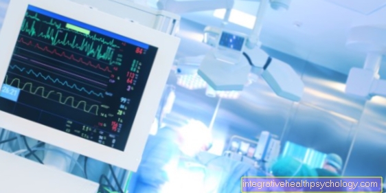
Monitoring is the term used to describe the monitoring of various circulatory parameters and physiological functions of a patient during an operation. Typically, the doctor in charge is an anesthetist. Depending on the type of operation, there are different forms of monitoring that can be expanded to include certain elements as required. In the following, basic monitoring, i.e. standard monitoring during an operation, will first be discussed.
Clinical observation
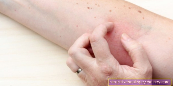
Nowadays patient monitoring is through modern technology very electronic become. However, the anesthetist should always do exactly that Keeping an eye on the patient to have. He pays particular attention to the even movement of the Rib cage of the patient, which is an indication of adequate Ventilation is. Also the Skin color of the patient can provide information about the success of ventilation, since, for example, with a Lack of oxygen the Lips blue can color. Furthermore, vegetative functions of the patient are observed, such as sweat, watery eyes and dilated pupils. These reactions can occur at insufficient depth of anesthesia occur.

- Ascending aorta -
Pars ascendens aortae - Aortic arch - Arcus aortae
- Thoracic aorta
(descending aorta) -
Thoracic aorta - Aortic slit of the diaphragm -
Aortic hiatus - Abdominal aorta
(descending aorta) -
Abdominal aorta - Aortic fork - Aortic bifurcation
- Trunk of the liver, spleen and ma
gene arteries - Celiac trunk - Upper arm artery -
Brachial artery - Common pelvic artery -
Common iliac artery - External head artery -
External carotid artery - Cervical artery (common head artery) -
Common carotid artery - Clavicle artery -
Subclavian artery - Axillary artery - Axillary artery
- Diaphragm - Diaphragm
- Renal artery - Renal artery
- Radial artery - Radial artery
- Ulnar artery - Ulnar artery
You can find an overview of all Dr-Gumpert images at: medical illustrations
Electrocardiogram (EKG)
The EKG draws the Cardiac current curve of the patient on. Do this to the patient Electrodes glued to extremities and chest. These then record potential differences caused by the electrical excitation line in the Heart occurrence. The anesthetist can use the ECG to determine the The speed of the heartbeat and the rhythm of the heart judge.
Blood pressure measurement
Of the Blood pressure is used in standard monitoring with the help of the so-called automatic non-invasive blood pressure measurement determined. To do this, a blood pressure cuff is placed on one extremity of the patient (usually on an arm). The cuff pumps up every 5 minutesso that the Vessels completely compressed. If the pressure is released again, arise Vibrationsas soon as that blood can flow again through the opening vessel. These vibrations are registered by the cuff. The maximum deflection of the vibrations corresponds to that mean blood pressure. This procedure will too oscillometric blood pressure measurement called. It is important that the Blood pressure cuff adapted to the patient is. Cuffs that are too small measure incorrectly high blood pressure values; cuffs that are too large measure incorrectly low values. The width of the cuff should be in about 2/3 the length of the upper arm correspond.
Oxygen saturation (SpO2)
To Monitoring of the oxygen content of the patient's blood is usually on one finger of one hand special clamp (Pulse oximeter) created. This clamp emits red light of different wavelengths. Because the blood depending on the oxygen saturation absorbs different wavelengths, the device can determine a saturation value from this. The norm is between 95 and 99%. However, the pulse oximeter can also display falsely high values, e.g. at a Carbon monoxide poisoning. With colored nail polish on the patient's nails, the values usually fall false low out. It also gives wrong values when the patient is in one State of shock because the blood is then increasingly shifted towards the center of the body.
Capnometry
The Capnometry denotes the measurement of the Carbon dioxide concentration in the exhaled air of the patient. The so-called end-tidal carbon dioxide (etCO2) measured what the CO2 is, what is completely at the end of the exhalation in the exhaled air. This is the most telling as it is the best CO2 concentration in the lungs reflects. At the beginning of the exhalation there is a higher proportion of oxygen in the exhaled air because the air is still from the windpipe is also exhaled that did not participate in the gas exchange in the lungs (so-called dead space volume).
The derivation of the capnometry in curve form is called capnography. It is particularly important because the CO2 values in the exhaled air can be used to check whether the ventilation tubes have been placed correctly. For example, if the CO2 level does not rise as soon as the patient is ventilated, this could indicate that the ventilation tube was inadvertently placed in the esophagus instead of the windpipe. In addition, a sudden and strong increase in the CO2 concentration can be an indication of so-called malignant hyperthermia, which can be a life-threatening reaction of the patient to certain narcotics.
Temperature measurement
The Measurement of body temperature is also an important part of monitoring. Typically the measurement in the nasopharynx or in the esophagus performed. This is important as the body is under anesthesia cool quickly can, because anesthetic agent the setpoint of the Body temperature adjust. This also explains what is often observed Shivering following a anesthesia. Children especially lose heat quickly. Therefore it is essential to have one Heat retention during the operation to pay attention.
Neuromuscular monitoring (relaxometry)
The neuromuscular monitoring of the patient is particularly important. So that Musculature The patient is usually relaxed during the operation and the doctors can easily perform the procedure so-called muscle relaxant administered. This medicine causes the Muscles temporarily paralyzed is. In relaxometry, the Effect and breakdown of these substances supervised. Two electrodes are used in the Distance of about 2-4 cm on the forearm of the patient glued, which is then directly over a there running annoy are located. By connecting the electrodes to a stimulator you can electrical impulses to stimulate the nerves be handed in. This leads to the corresponding muscle contracts (usually the Adductor pollicis muscle). As a result, the thumb of the patient. Based on the stimulus response, the Strength of the muscle relaxant used be assessed. This often happens according to certain patterns, for example the Train-of-four stimulation (TOF), where four electrical stimuli in a row and the extent of the stimulus response is recorded. Usually they fall Reactions gradually weaker out. If the blockage is very strong, there is no longer any response at all. Depending on the planned intervention, the desired neuromuscular blockade can be verified using this method. In addition, the relaxometry is also at Drainage of the anesthesia This is important because the ventilation tubes should only be removed when the relaxation of the muscles has largely been relieved, so that the patient can do so breathe independently again can.
Extended monitoring
The expansion of the basic monitoring can be indicated for certain interventions and patients. This applies in particular to patients with previous illnesses of the cardiovascular system or patients requiring intensive care.
Electroencephalogram (EEG)
The EEG records the brain waves. This gives clues to the Depth of anesthesia and blood flow to the brain. The EEG will in addition to normal monitoring used when particularly vulnerable people are operated on, or Interventions on blood vessels supplying the brain be made.
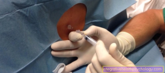
Invasive blood pressure measurement (IBP)
in the In contrast to non-invasive blood pressure measurement This procedure is more accurate because it is used to determine blood pressure Probe directly into an artery at the wrist is placed. This probe registered continuously check blood pressureso that even small fluctuations can be noticed immediately. The invasive blood pressure measurement is used especially for patients with Circulatory instability, if there is a high risk of bleeding or major vascular surgery.
Central venous catheter (CVC)
The central venous catheter represents an alternative access route to the patient's venous vascular system. It is usually placed in a large vein on the patient's neck. The central venous pressure can be determined through the CVC, which provides information about pressure conditions in the pulmonary circulation and thus indirectly about the heart function and the volume status of the patient. In addition, infusions and nutrient solutions can be given via the CVC, which would lead to irritation of the veins if the extremities were less accessible.
Extended EKG
In patients who are particularly at risk for one Undersupply of the heart muscle or a heart attack are, are through a more special EKG supervised. The so-called ST segment closely watched by the anesthetist recognize early can determine whether the patient's heart is receiving too little blood.
Measurement of cardiac output (CO)
The Cardiac output is the volume of blood that the heart pumps around the body in a given period of time. It is a Measure of the functionality of the heart muscle and is particularly reduced in the shock state. The cardiac output is measured using a so-called Thermodilution process. To do this, a temperature probe is inserted into a Vein in the groin placed. Then - usually in a vein in the neck area - a cold saline solution (approx. 20 ° C) injected. The distribution of the cold solution leads to a Change in temperature of the bloodwhich can also be measured in the bar after a certain time. The time it takes for the cold solution to be transported to the bar depends on cardiac output. This allows this to be calculated indirectly. This procedure is used especially for patients who are in shock, as well as in patients with sepsis (Blood poisoning).