Pituitary gland
Synonyms
Greek: pituitary gland
Latin: Glandula pituitaria
Pituitary gland anatomy
The pituitary gland is about the size of a pea and lies in the middle of the cranial fossa in a bony bulge, the sella turcica (Turkish saddle, with a shape reminiscent of a saddle). It belongs to the diencephalon and is located in close proximity to the junction of the optic nerves. It is only separated from the nasopharynx and the sphenoid sinus, a paranasal sinus, by the bony base of the skull. The pituitary gland is connected to the hypothalamus above it via the pituitary stalk (infundibulum).
The pituitary gland is anatomically divided into two parts: the anterior pituitary gland (Adenohypophysis) and the posterior pituitary gland (Neurohypophysis). These two parts developed from different parts. While the anterior pituitary produces its own hormones, the posterior pituitary only releases the hormones produced by the hypothalamus, to which it is connected via small blood vessels.

Cerebrum (1st - 6th) = endbrain -
Telencephalon (Cerembrum)
- Frontal lobe - Frontal lobe
- Parietal lobe - Parietal lobe
- Occipital lobe -
Occipital lobe - Temporal lobe -
Temporal lobe - Bar - Corpus callosum
- Lateral ventricle -
Lateral ventricle - Midbrain - Mesencephalon
Diencephalon (8th and 9th) -
Diencephalon - Pituitary gland - Hypophysis
- Third ventricle -
Ventriculus tertius - Bridge - Pons
- Cerebellum - Cerebellum
- Midbrain aquifer -
Aqueductus mesencephali - Fourth ventricle - Ventriculus quartus
- Cerebellar hemisphere - Hemispherium cerebelli
- Elongated Mark -
Myelencephalon (Medulla oblongata) - Big cistern -
Cisterna cerebellomedullaris posterior - Central canal (of the spinal cord) -
Central canal - Spinal cord - Medulla spinalis
- External cerebral water space -
Subarachnoid space
(leptomeningeum) - Optic nerve - Optic nerve
Forebrain (Prosencephalon)
= Cerebrum + diencephalon
(1.-6. + 8.-9.)
Hindbrain (Metencephalon)
= Bridge + cerebellum (10th + 11th)
Hindbrain (Rhombencephalon)
= Bridge + cerebellum + elongated medulla
(10. + 11. + 15)
Brain stem (Truncus encephali)
= Midbrain + bridge + elongated medulla
(7. + 10. + 15.)
You can find an overview of all Dr-Gumpert images at: medical illustrations
function
The pituitary gland is a hormonal gland that is part of the endocrine system. It has an overriding control function in the hormonal balance.
The regulation of the hormonal balance of a person is very complex and comprises three levels of control: we have the as the top regulatory unit Hypothalamus. This pours Liberine and Inhibins control hormones that induce the pituitary gland to release hormones in turn.
The pituitary gland can be described as the second highest regulatory unit. She in turn pours stimulating Hormones from that Tropinethat act on the body's hormonal glands.
These glands, roughly thyroid, Testicles, Ovary and Adrenal cortex, are the third institution to release the free hormones. These free hormones directly influence the body, water, sexual and energy balance.
The following hormones are produced in the anterior lobe of the pituitary gland: TSH (Thyrotropin), LH (Luteinizing hormone), FSH (Follicle-stimulating hormone), STH (Somatotropin, also GH for English Growth Hormone), ACTH (Coticotropin), MSH (Melanotropin) as well Prolactin.
The TSH formed in the pituitary gland is the thyroid-stimulating hormone. It stimulates their growth and promotes the secretion of thyroid hormones from the thyroid gland.
LH and FSH are both at man as well as the woman important hormones that control the sexual balance. LH solves that ovulation of the woman, promotes growth and education of the for the pregnancy important corpus luteum. In men, LH promotes this Testosterone production in the testicle. FSH promotes the maturation of women ova in the ovary, in men the maturation of the Sperm cells.
GH or STH is important for that growth of all organs as well as the growth in length of the trunk and arms and legs. It is released to a greater extent in childhood during growth spurts, but it is still a necessary growth hormone in adults.
ACTH stimulates the adrenal cortex in response to this stimulus in particular cortisone forms. This is one Stress hormonewhich is important for the Blood sugar increase, the suppression of excessive inflammatory reactions, the protein turnover and much more.
The MSH of the pituitary gland stimulates the Pigment cells (Melanocytes) of the skin Color pigmentation.
Prolactin is the hormone that helps the mammary gland of a pregnant or breastfeeding woman to grow and Milk production stimulates.
The following hormones are produced in the posterior pituitary gland: Oxytocin and ADH (Antidiuretic hormone or adiuretin or vasopressin).
Oxytocin is a hormone with a variety of functions. It is also called "Cuddle hormone“Because it is released upon physical contact. It is also important for training Labor pains under the birth. Ultimately it will be the Breastfeeding and leads to milk being released towards the nipple.
ADH is a hormone that is involved in regulating the water balance. It promotes the reabsorption of free water in the Kidneysso that less water is excreted with the urine and consequently the Blood pressure increases.
Pituitary gland disorders
Pituitary Insufficiency
Synonyms: hypophysis, hypopituitarism
Inflammation, injuries, radiation or bleeding can cause disorders in the pituitary gland. This can result in the production of both the posterior lobe of the pituitary and the anterior pituitary hormones. Most hormonal failures occur in a coupled manner. So it is either all hormones of the anterior lobe (anterior pituitary insufficiency), those of the posterior lobe (posterior lobe of pituitary insufficiency) or all hormones are reduced at the same time. The consequence is that the downstream hormone systems release fewer hormones, which results in disorders of the corresponding body function.
Symptoms of hypofunction of the pituitary gland can be, for example, short stature with a lack of STH, menstrual disorders and lack of expression of the sexual organs during puberty with a lack of LH and FSH, drop in blood pressure and greatly increased water excretion with ADH deficiency.
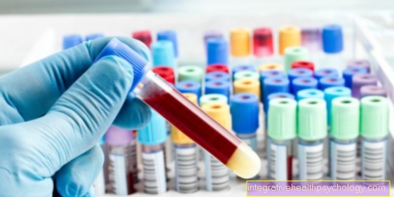
To establish the diagnosis, the hormone levels are determined by taking a blood sample and CT or MRI of the skull are made.
The therapy of hypofunction of the pituitary gland consists in the drug administration of the missing hormones.
Pituitary adenoma
Benign growths can appear in the front part of the pituitary gland. These are known as adenomas. Usually these adenomas produce hormones which are then present in increased amounts in the blood.
The pituitary adenoma is divided into a microadenoma (smaller than 1cm) and a marrowadenoma (larger than 1cm).
The most common benign tumor of the pituitary gland is the prolactinoma, a tumor that produces prolactin. The consequences are breast growth and milk running even without pregnancy.
STH-producing tumors lead to tall stature before the end of their growth and after puberty to acromegaly, a disease in which the fingers, nose, mouth, tongue and ears thicken considerably.
ACTH-producing tumors stimulate the adrenal cortex to produce cortisol and lead to Cushing's disease with truncal obesity, full moon face, muscle breakdown, high blood pressure, susceptibility to infection and high blood sugar.
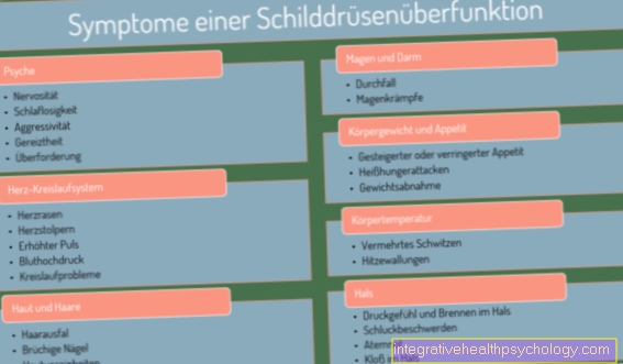
TSH-producing adenomas cause an overactive thyroid with sweating, a racing heart and weight loss.
Pituitary gland tumors can lead to headaches and pressure on the optic nerve junction, which can result in blinker blindness.
These hormone-forming tumors are detected by detecting elevated levels of hormones in the blood. If all values are normal, a non-hormone-producing adenoma may still be present. Imaging with CT or MRI must also be performed.
Due to their anatomical location, pituitary gland tumors are usually operated on via what is known as a transsphenoidal approach. The surgeon has the option of removing the overgrown part of the pituitary gland without visible scarring via the nose, the paranasal sinus behind it and by breaking through the thin bony floor under the pituitary gland.
If an operation is not possible or desired, there are drugs to suppress hormone production.
Also read the article: Cushing's disease.









.jpg)

