Neurofibromatosis type 2
Note
You are currently on the start page of the topic Neurofibromatosis type 2.
On our other pages you will find information on the following topics:
- Symptoms of neurofibromatosis type 2
- Neurofibromatosis type 1
- Symptoms of neurofibromatosis type 1
- Life expectancy and therapy for neurofibromatosis type 1
Classification of neurofibromatoses
- Neurofibromatosis type 1
and - Neurofibromatosis type 2
Synonyms
NF2
definition
Type 2 neurofibromatosis is like that NF 1 a hereditary disease from autosomal dominant inheritance. This means that if an NF gene is present (there is always a gene from mother and father), the disease occurs.
The genetic mutation lies on Chromosome 22q12.2.
It is about a Tumor disease. The Tumors can be found in the Nervous systemi.e. especially along the Cranial and spinal nerves (spinal cord nerves).
Epidemiology / incidence in the population
The Type 2 neurofibromatosis With an incidence of 1: 25,000 to 35,000, it occurs significantly less than NF1.
In this condition are Men and Women equal often affected.
Half of the cases are new mutations, i.e. not inherited cases.
Etiology / cause

The gene locus of the NF2 gene is on chromosome 22q12.2. The gene codes for the protein Merlin. Merlin is also called Neurofibromin 2 or Schwannomin. The task of this protein is to anchor the supporting skeleton of the cells, here the actin cytoskeleton, in the cell membrane, especially that of the nerve cells.
Another task is to inhibit the transmission and amplification of extracellular signals (signals outside the cell) that are transmitted by growth factors.
A mutation means that signals that stimulate the cell to divide are not inhibited, which leads to increased cell division. Since NF2 is one of the tumor suppressor genes (tumor protection genes), a mutation of this gene promotes tumor development.
Finally, Merlin is involved in the regulation of cell adhesion, i.e. the connection between cells.
More information on neurocutaneous syndromes can be found here: Neurocutaneous Syndrome
Complications
Since the Tumors occur along the course of the nerves, depending on the location and function of the affected nerve, there is a weakening or even complete loss of function.
- deafness
- Loss of vision or poor eyesight
and - Paralysis
are among the most common complications.
Even benign tumors are always at risk of malignant degeneration.
diagnosis
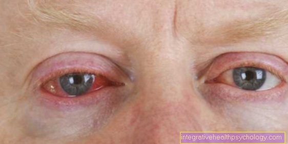
Lens opacities in childhood are atypical, so this usually the first and very early symptom should always be thought of as neurofibromatosis type 2. Affected by increasing Loss of vision and one Glare sensitivity noticeable.
Please also read our topic: Symptoms of neurofibromatosis type 2.
Of the progressive hearing loss usually begins years before the diagnosis.
As with NF1, there are also clinical ones Diagnostic criteria.
The detection of bilateral tumors of the auditory and equilibrium nerves using imaging methods is a clinical diagnostic criterion.
If a patient has first-degree relatives with a confirmed diagnosis of neurofibromatosis type 2 and they experience early lens opacities or Neurinomas, neurofibromas, meningiomas or Gliomas occur, this is considered a further clinical diagnostic criterion.
Laboratory analytical methods for the detection of the mutated gene are possible, but at the same time both expensive and time-consuming.
- Imaging procedures (X-ray, CT, MRT)
- Hearing test (audiometry)
and - Functional tests of nerves (e.g. acoustic evoked potentials AEPs)
are the means of choice, especially to determine the severity or progression of the disease.
Features, symptoms and ailmentsAge
Type 2 neurofibromatosis typically manifests between the ages of 18 and 24 years.
Lens opacities
However, the so-called "Subcapsular posterior cataracts". This is a special form of lens opacity comparable to that Grey star in old age.
Nervous system tumors
In the Neurofibromatosis type 2 it is a tumor disease that the Nervous system concerns. Typically found in these patients Meningiomasi.e. Meningen tumors and neuromas.
Neuromas, or also Schwannomas called, are benign tumors that arise from Schwann cells.
The task of Schwann cells is to enclose nerve fibers, to protect them, to isolate them along the long branches and thus to enable their function. The multiplication of the Schwann cells leads to a functional restriction or a functional failure of the affected nerves.
In 80% of those affected, these schwannomas develop on both sides along the 8. Cranial nerve.
Since this Vestibulocochlear nerve is responsible for hearing and balance, symptoms arise as progressive Hearing loss to deafness, gait disorders and equilibrium and, as a result, noises in the ears (Tinnitus) and dizziness.
Only one side is affected in about 6% of patients.
Other cranial and peripheral nerves can also be affected.
Neurofibromas
The clinical similarity to neurofibromatosis type 1 arises when, when subcutaneous, i.e. Peripheral nerves located in the subcutaneous fat tissue are affected, which then appear like neurofibromas. Histologically, that means fine tissue, there is no similarity.
About half of those affected show Café-au-lait spots. Rarely do more than 3 spots appear.
therapy
Since the Neurofibromatosis type 2 If it is a genetic disease, therapy to eliminate the cause is not possible.
Therefore, the Therapy according to the symptoms.
By means of surgical procedures on the eye, it is now possible to remove cloudy lenses artificial lenses to replace.
In order to avoid progressive hearing loss, it is recommended that tumors of the Auditory and equilibrium nerves to be surgically removed at an early stage.
If other cranial or spinal nerves are also affected, an operation is recommended in order to maintain the residual function of the nerves. But such operations also harbor dangers. Nerves can be damaged by the operation. In addition, in not uncommon cases the Tumors again.
Therefore, regular preventive and check-ups should take place. One should also keep in mind that benign tumors always pose a risk of malignant degeneration.
Is the Hearing loss to deafness advanced, one should consider the possibilities of Cochlear or brain stem implants use.
Electrodes are implanted in the inner ear or brain and the affected person can hear again or take part in far-reaching communication.











.jpg)

