What is pleural empyema?
Definition - what is pleural empyema?
The translation of the term "pleural empyema" means an accumulation of pus in the pleura.
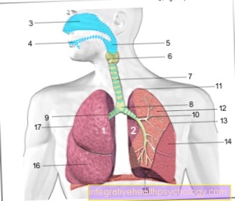
The pleura describes a covering of the lungs, which consists of two leaves. The lungs themselves are covered by a thin sheet of pleura, the so-called "pleura visceralis". From the outside, the "parietal pleura" rests on it, which is attached to the chest wall and the inner ribs. In between there is a cavity in which there is a negative pressure and small amounts of a lubricating fluid. The undamaged negative pressure in the pleura enables the lungs to unfold and thus the inhalation and exhalation processes.
Pus in this area suggests inflammation, which can develop into dangerous courses due to the obstruction of breathing and the proximity to vital chest organs.
In order to understand the exact development of the pleural empyema, read in advance general information about empyema and the structure of the pleura. The following articles promise to provide an overview:
- Structure of the pleura
- What is an empyema?

I recognize pleural empyema by these symptoms
The pleural empyema is only a consequence of the underlying inflammation and previous illnesses. As a rule, the underlying diseases cause symptoms that also suggest pleural empyema.
These include above all coughing with expectoration, shortness of breath, rattle noises when breathing, high fever, chest pain and a greatly reduced general condition. These are primarily symptoms of acute pneumonia, which is the most common cause of pleural empyema.
The empyema itself can exacerbate chest pain and shortness of breath, as the increased fluid build-up around the lungs can restrict the expansion of the lungs and thus impede breathing.
A slight accumulation of pus after the pneumonia has subsided, however, can also be symptom-free.
Since pleural empyema is very much associated with pneumonia and the symptoms are very similar, it is advisable to take a look at the following page: What are the symptoms of pneumonia?
What do pleural empyema and pleural effusion have in common?
Pleural effusion is the term used to describe all accumulations of fluid between the two sheets of the pleura. In addition to pus, this can primarily be water and blood, which can be attributed to numerous causes.
Empyema is only a relatively rare form of pleural effusion. The most common cause of fluid build-up in the pleura is heart failure.
Such effusions can be recognized in the X-ray image from an amount of 250 ml. Punctures of the effusion are only rarely necessary, as the fluid can in many cases be absorbed by itself.
An overview with the most important information about pleural effusion can be found at:
- What is a pleural effusion?
- Water In Your Lungs - What To Do About It
Course of disease in pleural empyema
Pleural empyema is usually preceded by an infectious disease that can enter the body in various ways. Only when this inflammation reaches the edges of the lungs and the pleura can pus accumulate there. The original inflammation can be highly acute and active, or it can already be largely healed or it can be encapsulated as an abscess.For this reason, the course, duration and prognosis of pleural empyema vary greatly.
With the intake of antibiotics, the empyema usually improves within 2-3 weeks. The empyema can partially self-resorb.
Therapeutically, however, in almost all cases of an infectious effusion, a drain is inserted in the chest, which sucks out the pus immediately. The amount of pus that is sucked off slowly decreases in the course of the therapy until there is no more effusion.
What to expect if there is pus in the lungs? The process and possible complications can be found in the following article: What happens if there is pus in the lungs?
Duration of a pleural empyema
Pleural empyema only develops if there is still active inflammation in the surrounding tissue. In most cases it is an acute pneumonia.
The duration of the illness can vary widely and depends on many factors. The correct therapy and the response of the immune system are crucial.
Harmless pneumonia will usually clear up within 2-3 weeks. The formation of pus in the pleura should also subside within this period.
In severe cases, if the therapy does not respond, the healing time can be delayed indefinitely.
Since the presence of pleural empyema depends on the presence of active inflammation, it makes sense to consider the duration of the inflammation. You can find the most important information on this at: How long does pneumonia last?
Treatment of pleural empyema
The treatment of the underlying disease is in the foreground. If there is an inflammation in the chest area, it must be treated with antibiotics, surgery or other measures as soon as possible before it can spread to surrounding organs.
The pleural empyema is also treated with antibiotics in most cases. In addition, a tube is usually placed between the pleural leaves to aspirate the empyema. This remains as a drainage in the chest to be able to suck out further pus formations until the inflammation has subsided. Smaller effusions resorb by themselves. Surgical treatment of the inflammation is rarely necessary.
The prevention of previous illnesses, i.e. usually pneumonia, can also be of interest to you. You can find out more at: How helpful is vaccination against pneumonia?
When do you need surgery with pleural empyema?
The installation of a drainage between the pleural leaves is only a slightly invasive therapeutic intervention.
Only in rare forms of peural empyema does an actual surgical opening of the chest need to be performed. This is necessary with encapsulated inflammation and special locations of the pleural empyema. For this purpose, a small fenestration of the ribs can be made over the site of the inflammation in order to clear out the inflammation, to rinse and to insert a drainage.
In the case of severe and long-lasting pleural empyema, the pleural leaf can also be removed or a sponge containing antibiotics can be inserted.
At this point, you can also read the various types of drainage to discover the most suitable method for your pleural empyema: What is a chest tube?
Puncture for a pleural empyema
In most cases of pleural empyema, both diagnostic and therapeutic punctures are required.
Diagnostic puncture is the most common method for reliably detecting a collection of pus and examining the sample in the laboratory for the exact pathogen.
In most cases, a puncture of the pleura must also be performed therapeutically in order to drain the pus and to insert a drain that can drain away future pus.
The puncture is usually performed under local anesthesia and sterile in order not to introduce any further pathogens into the chest.
In everyday clinical practice, a pleural puncture is not uncommon and promises, with a low risk, a detailed diagnosis of the underlying disease and relief of the lungs. Everything you need to know about "pleural puncture" is explained below: How is a pleural puncture performed?
Causes of pleural empyema
The most common cause of pleural empyema is bacterial inflammation of the leaves of the lung membrane.
Normally, the pleura is a closed space that neither air nor bacteria can reach. Bacteria can only trigger inflammation there if they get to one of the leaves of the pleura from the outside or inside, for example via the lungs. Most often this happens in the context of pneumonia. If the inflamed area of the lung is close to the pleural leaf resting on the lung, the inflammation can spread into the pleural cavity. The infected area can produce a purulent inflammatory secretion, which is emptied between the pleural leaves and leads to an effusion in the pleura called "empyema".
Other ways in which pathogens can be introduced into the pleura are blood poisoning, a previous operation, a tear in the esophagus or an external injury, for example through a stab wound in the chest area.
Find out more about the topic on our main page: Pleurisy - what's behind it?
Lung abscess as the cause of pleural empyema
A lung abscess is an enclosed area in the lungs that contains inflamed tissue. The abscess is often a consequence of acute pneumonia, but does not itself lead to an acute clinical picture.
The lung abscess only becomes dangerous if parts of the encapsulated inflammation spread to the rest of the lungs, the airways or the pleura. The latter can lead to pleural empyema as a result.
Pneumonia can lead to a lung abscess, which can lead to pleural empyema. Study the individual clinical pictures in detail to prevent this vicious circle. The most important information on the diseases can be found at:
- Lung abscess - what's behind it?
- What is pneumonia?
Pneumonia as a cause of pulmonary empyema
Pneumonia is a very common disease that can be a dangerous disease, especially in immunocompromised, old or very young people.
As a rule, pneumonia can be caused by any bacterial, viral, parasitic or other pathogen, with bacterial and viral infections being the most common and causing the most acute courses. If pneumonia spreads quickly in the chest area, it can spread to other organs, affect the pleura and even cause life-threatening blood poisoning.
Pleural empyema is therefore a common complication of acute pneumonia.
In order to avoid such complications, it is very important to identify pneumonia immediately and to initiate appropriate treatment. For this reason, the following article also plays an important role: Signs of pneumonia
How contagious is pleural empyema?
In principle, pleural empyema and its underlying disease are contagious.
In most cases, however, the pleural empyema is encapsulated in the chest and therefore represents a negligible risk of infection.
However, underlying pneumonia can be contagious, depending on the pathogen. Pathogens can be distributed in particular via the cough secretion, which can lead to infections, especially in the case of immunocompromised or particularly contagious pathogens, for example tuberculosis.
The following article also provides information on whether and how you should protect your fellow human beings in the event of pleural empyema and the underlying pneumonia: How contagious is pneumonia?
Diagnosis of pulmonary empyema
The diagnosis begins with a detailed questioning and physical examination of the patient. Pleural empyema is usually preceded by previous lung diseases or serious external injuries, which can be determined with the help of a survey. Inflammation of the lung tissue is often externally recognizable as a cough, shortness of breath and fever. During the physical examination, weakened breathing sounds and muffled knocking noises may be heard on the chest.
The collection of pus can be recognized diagnostically on a radiological image, for example an X-ray or computer tomography.
In order to identify the effusion in the pleura as an accumulation of pus and, if necessary, to determine a precise pathogen, the effusion can be suctioned off with a puncture needle and examined more closely in the laboratory.
You might also be interested in these topics:
- CT of the lungs
- Puncture - what's behind it?
What does pleural empyema look like on X-rays?
The X-ray is the first diagnostic measure after taking the medical history and the physical examination. The X-ray shows coarse types of tissue in the chest area, so that it is possible to differentiate between air, fluids, organs and bones.
For all lung diseases, an X-ray can provide information about the density of the lung tissue, possible inflammation and fluid accumulation in the lungs or between the pleura leaves.
In the case of pleural empyema, an accumulation of fluid in the form of a decrease in the transparency of the image can often be recognized at the lower edges of the lungs. The fluid collects on the X-ray images due to gravity in the lower area near the diaphragm.
On the basis of the X-ray image, however, it cannot be differentiated which type of effusion is between the pleural leaves and where the effusion comes from.
A sample of the liquid must therefore be obtained for more detailed diagnosis.
Recommendation from the editor
Further information that may be of interest to you:
- What is meant by empyema?
- Life expectancy with water in the lungs
- Diseases of the lungs
- Pneumonia without a cough
- Pneumonia without a fever


.jpg)


