Abdomen
definition
The abdominal cavity, also known as the abdominal cavity, begins below the diaphragm and extends to the level of the iliac crest. There the abdominal cavity merges into the smaller pelvic cavity, which extends to the pelvic floor. The entire abdominal and pelvic cavity is lined by the peritoneum. From the outside, the abdominal area is stabilized and protected by the abdominal muscles. The upper part is also surrounded by ribs. The abdomen is home to a large number of organs. In addition to the digestive organs, it also contains the spleen.

Abdominal anatomy
The organs are not free in the abdominal cavity, but are about the so-called Mesentery connected to the abdominal wall. The mesentery is a Peritoneum fold on the back of the abdominal wall, through the blood vessels to the individual organs.
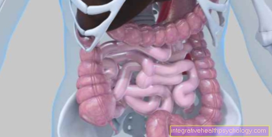
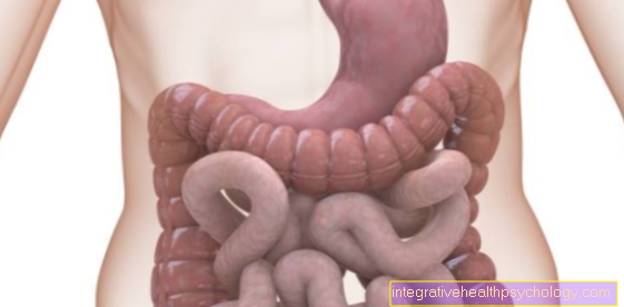
The organs in the abdomen include the stomach, the liver, the Gallbladder, the pancreas, of the Thin- and Large intestine and the spleen (The exact location of the organs is discussed below). The Kidneys are not in the abdomen, but behind it, built into one Fat capsule. In the pelvic cavity are the bladder and the internal genital organs, by the man prostate, with the woman the Ovaries and the uterus. In addition to the organs, the abdominal cavity is also traversed by the large vessels (the aorta and the Inferior vena cava), which supply the abdominal organs and then pull them further to the legs. Besides, there is also still Adipose tissue in the abdominal cavity. The structure of the peritoneum which is interspersed with a lot of fatty tissue and lies in front of the small intestine should be emphasized.
Right abdomen
The upper right abdomen is covered by the liver with the small gallbladder filled out. The gallbladder is almost completely covered by the liver. The liver can be thought of as a triangular structure, one leg of which pulls into the left abdominal cavity and the other downward. In some diseases, the liver can become so large that it fills a large space in the abdominal cavity and extends far into the lower right abdominal cavity.
In the middle of the upper abdomen is the transition from stomach to the Duodenum, also Duodenum called. Behind it lies the head of the pancreas. Access via two execution corridors bile from the gallbladder and Pancreatic secretions ins Duodenum. This is particularly important for fat digestion. Below these organs, the colon moves once from the right to the left side. The transition from small intestine to large intestine is located in the lower right abdomen. At this point is the appendix with his Appendix to find a pain that causes inflammation.
You can find more information about the anatomy of the abdominal cavity here: Digestive tract
Left abdomen
The first is found in the upper left abdomen stomachthat is in the middle of the abdomen in the Duodenum transforms. Hidden behind the stomach is the pancreas, the head of which is surrounded by the duodenum. Her tail pulls back to the left to the spleen. The pancreas also produces digestive secretions insulin, which is necessary for a stable blood sugar level. The spleen is in the Spleen niche hidden, which lies deep behind by the ribs. In the case of broken ribs, however, it can be injured by this, so that it becomes a Rupture of the spleen and internal bleeding can occur. Below the spleen is the kidney With Adrenal gland embedded in their fat capsule. In the lower abdomen are as on the right Thick- and Small intestine. The small intestine is 5-6 meters long and takes up a lot of space. It is surrounded by the large intestine like a frame. This begins in the right lower abdomen and moves in the left lower abdomen into the pelvic cavity, where it merges into the rectum.
Lymph nodes in the abdomen
The lymphatic system, a fluid system that exists parallel to the blood vessels, is particularly useful for the body's immune defense. The filtered, excess tissue fluid is filtered and channeled through several large and small lymph nodes to the large veins in the upper chest area. In the abdomen, the lymph nodes can be divided into superficial and deep lymph nodes, with the former leading into the deep ones. By definition, the superficial lymph nodes do not belong directly to the abdominal cavity.The deep lymph nodes follow the course of the abdominal aorta, i.e. the largest artery in the abdomen. From most abdominal organs, with the exception of the descending large intestine, the lymph first flows into a trunk of the internal organs and is collected there and sent on to the lymphatic drainage of the upper body. On the way there are the abdominal cavity lymph nodes and the mesenteric lymph nodes. The lower organs drain the lymph through the inguinal lymph nodes. These also lead in the further course into the lymphatic system of the upper body. For each organ, a so-called sentinel lymph node can be named, which is the first lymph node to be infected in the case of spreading carcinomas. This is always removed and pathologically examined during tumor operations.
Abdominal pain
The body projects pain in the individual abdominal organs onto certain skin areas so that an assignment can be possible. Pain of pancreas are perceived as a belt on the back below the shoulder blades and in the middle of the upper abdomen.
During the stomach leads to pain in the left upper abdomen, it is with the liver and the Gallbladder the right upper abdomen. The gallbladder can also cause pain in the right shoulder. Small intestine and Large intestine lead to pain in the middle abdomen. At a Appendicitis pain in the right lower abdomen is characteristic.
In addition to the places of pain perception, one can also Pain qualities distinguish. For one thing, there is colicky pain that makes it difficult to lie still. They appear in waves. The pain can be compared to a muscle spasm that does not want to go away. The cause is usually with Gall or kidney stonesobstructing the bile duct or ureter. In the case of inflammation in the abdominal cavity, e.g. appendicitis or Inflammation of the gallbladder, the pain is rather dull and increases with tremors. Because of this, those affected usually lie still.
Burning sensation in the abdomen
The statement that there is a burn in the stomach is a description of a certain type of pain. Burning usually represents a rather continuous form of pain and thus points to inflammatory processes and not to an acute injury or stone disease. Inflammation can affect all organs and surrounding tissues. Common inflammations are appendicitis and diverticitis. Peritonitis and pancreatitis are less common, but much more dangerous. Burning pain in the abdominal area can also occur with inflammation of the bladder and kidney pelvis. An important differential diagnosis in burning epigastric pain is acute posterior wall infarction. This is a heart attack, the pain of which radiates into the abdomen rather than the left arm. Acute, severe pain in the abdominal area should always be clarified by a doctor, as the causes range from simple gastrointestinal flu to life-threatening peritonitis.
What does air in the abdomen mean?
In a healthy person, there is no air in the abdomen, except in a hollow organ such as the intestine. Air outside hollow organs is called free air. This can be in the abdominal cavity for several days after operations and then has no disease value. Otherwise, free air is an indication of a breakthrough of hollow organs (perforation). The contents of the hollow organ exit through an opening into the abdominal cavity, i.e. air, digestive juices, chyme or stool. This leads to dangerous inflammation in the abdomen and severe pain.
There can be many causes for a perforation. Such injuries can result from blunt abdominal trauma in accidents or from stab wounds. But acute or chronic inflammatory processes and tumors can also lead to perforations. Free air in the abdomen is usually diagnosed using X-rays. However, free air cannot be detected in the X-ray with every perforation. The exact cause must be determined in further examinations. As a rule, the cause must be treated as quickly as possible.
Read our topic: Air in the abdomen
Adhesions in the abdomen
Abdominal adhesions that too Adhesions often arise between the peritoneum and the Serosa, a skin that covers the abdomen. Often the cause of adhesions lies in operations, after which the tissue heals again and in some cases scars. Minimally invasive procedures, such as a laparoscopy, result in fewer adhesions. But inflammation in the abdomen can also be the cause.
Usually adhesions do not lead to any complaints. However, it is possible that they restrict the mobility of the bowel or even pinch the bowel, which requires emergency surgery. In the long term, recurring pain and irregular stool can occur. In the event of serious problems, the adhesions must be loosened. This is usually done in a laparoscopy to keep the risk of new problematic adhesions low. Existing adhesions are also loosened when the operation is repeated. By cutting through the adhesions, however, small wounds arise, which can lead to adhesions again during the healing process.
In general, the surgical loosening of adhesions does not have a good chance of success, so that complaints caused by adhesions are normally not treated surgically. One tries to alleviate the symptoms with drugs or alternative therapies.
Further information on this topic can be found on our website: Adhesions in the abdomen
Free fluid in the abdomen
The free fluid in the abdomen can be different fluids. Blood, pus, wound fluid, urine and porridge are possible. Which liquid is involved depends on the cause. Urine is found in accidental damage to the bladder or in the event of a leak in the urinary tract after operations. Mashed food is also conceivable for organ leaks due to accidents or serious infections. Wound fluid and pus are usually the result of inflammation and infection in the abdomen. If an inflamed appendix or diverticulum ruptures, these can get into the abdomen and cause severe peritonitis.
Blood gets into the abdomen especially in accidents. Traffic accidents can tear large organs with good blood circulation, such as the spleen, and bleed into the abdomen. Even if an aneurysm tears, a lot of blood will get into the abdomen. Free fluid in the abdominal cavity always has a disease value and requires treatment. In the event of severe injuries, an examination for free fluid in the abdominal cavity is carried out in the emergency room's emergency room. This allows conclusions to be drawn about internal injuries. The liquid always collects at the lowest point. When standing this is the pelvis and when lying down an area near the kidneys.
Causes of blood in the abdomen
Basically, all blood in the human body is intravascular, i.e. within blood vessels. If blood gets into the abdominal cavity, this always has a disease value.
One cause of blood in the abdomen is cracks in the abdominal organs. This can occur due to inflammation or accidents. In the case of inflammation in the abdominal cavity, such as appendicitis or diverticitis, vessels can be damaged and if the affected organ ruptures, pus and wound fluid can also transport blood into the abdomen. With such ruptures there is a fundamental danger to life. In the case of traffic accidents, organs can also tear, regardless of previous damage. The liver and the spleen, both of which are very well supplied with blood, are particularly at risk.
Large vessels can also tear directly if they are exposed to strong forces. People with previous vessel damage can also develop protuberances on the vessel wall, so-called aneurysms. These can tear independently of an accident and are a direct indication for surgery. In this case, many of those affected cannot make it to a clinic in time. In women, an additional cause of abdominal bleeding is endometriosis or an abdominal pregnancy. In the first case, it is the lining of the uterus outside the uterus, which is broken down depending on the cycle. Abdominal pregnancy is an embryo that implants outside of the uterus and can infiltrate large blood vessels. Acute abdominal bleeding is always an emergency.
Also read: Rupture of the spleen
Tumors in the abdomen
Tumors are generally classified according to their cell type and malignancy. Many tumors are caused by glandular tissue, which is also found in many places in the abdomen. If they are malignant, these are called carcinomas. Benign gland tumors are called adenomas. Malignant tumors from muscle cells or connective tissue are sarcomas. Both benign and malignant tumors can develop in all abdominal organs and in between.
Classic gastric carcinoma starts from the mucous membrane cells of the stomach and can be traced back to an infection with the Helicobacter pylori bacterium. Benign protrusions of the mucous membrane called polyps can also occur in the stomach. The liver can also be affected by carcinoma. These usually arise as a result of a change in the structure of the liver in the event of infections or from high alcohol consumption. The pancreas is also a source of cancer, which is often discovered late. Colon cancer is a very common cancer in Germany. This often affects the rectum and can be detected early through regular preventive measures. Colon polyps are also common. In addition to the so-called primary tumors, metastases from other organs can also spread into the abdomen.
Both carcinomas and all other tumors can also affect the peritoneum and other structures between the organs. By definition, kidney tumors do not count among the abdominal cavity tumors, as the kidneys are located behind the peritoneum, or retroperitoneally in technical terms. Benign abdominal tumors can also be dangerous if they restrict other structures. This is conceivable in the pancreas, for example, since a benign tumor can prevent digestive juices from draining. Masses in the abdominal cavity are therefore always worth observing and often also require treatment.
More about this at: Tumor in the abdomen
Metastases in the abdomen
Daughter tumors (medical Metastases can be found anywhere in the abdomen. Many tumors of the abdominal organs metastasize to the liver through the bloodstream. This is because venous blood from the digestive organs flows through the liver before returning to the heart. Tumors can also spread over the lymphatic vessels, allowing metastases to form in lymph nodes. One differentiates whether regional or lymph nodes remote from the tumor are affected. The regional lymph nodes are lymph drainage stations of the affected organ and are also removed during surgical treatment of the tumor.
Please also read our topic: Peritoneal cancer
Lipoma in the abdomen
A lipoma is a benign tumor that arises from adipose tissue. This tumor can be of any size and is usually clearly demarcated and even movable in relation to surrounding structures. The so-called omentum majus, an apron made of fatty tissue that protects the abdominal organs, is located in the abdomen. From this lipomas can arise. There are also small fat appendages on the large intestine, which can increase in size unnaturally. As such, lipoma is not a hazard and there is no need for treatment. However, as soon as the lipoma narrows other structures in the abdominal cavity, surgical removal must be considered. This can be necessary if there is pressure on vessels or nerves or on the intestines. With a rapidly growing lipoma or an unusual size, as well as in the absence of delimitation from the tissue, further diagnostics, for example computed tomography, should be carried out in order to rule out a malignant liposarcoma. Lipomas in the abdomen are relatively rare. Most lipomas are found in the subcutaneous fatty tissue of the arms and legs.
You might also be interested in this topic: When should a lipoma be removed?
Abdominal lymphoma
A lymphoma is a malignant neoplasm that arises from lymphatic cells. These can be cells of the bone marrow, the spleen or other organs of the immune system. Lymphoma cells can settle with the blood throughout the body and thus also in the abdomen. Primary lymphomas can develop in the abdomen, for example in the spleen or in certain areas of the intestine. The prognosis for lymphoma depends on many different factors, such as age, the stage of the disease, and the type of lymphoma.
Also read: Treatment of lymphoma
Cyst in the abdomen
Cysts are spherical, fluid-filled cavities that can occur in almost all organs. Small cysts, for example in the liver or ovaries, do not require treatment and do not cause any symptoms. Larger cysts should be monitored regularly on ultrasound so that an increase in size can be detected. If an organ is affected by many cysts, the organ function can be restricted. In this case, the cyst must be removed. Pressure on other structures can also cause pain or restrictions and removal of the cyst may be useful. In rare cases, cysts can also become malignant, which is why the controls are useful. The cause of cysts varies widely. Ovarian cysts are often hormone-related. Chronic diseases such as cystic fibrosis or tumors can also cause cysts to form. Since most cysts are uncomfortable, these are often incidental diagnoses when an abdominal imaging is done for other reasons. Cysts can be seen on ultrasound, computed tomography or MRI.
Testicles in the abdomen
During embryonic development, the testicles are located in the abdomen and only at the end of pregnancy do they migrate down into the scrotum. In some babies, especially those born prematurely, this development is not quite complete. By the age of one, the testicles should finally have moved into the scrotum. If this is not the case, the boys have to undergo an operation, since overheating of the testicles limits fertility and can also lead to degeneration. The position of the testicles is checked by the pediatrician during the preventive examinations and a referral to the pediatric urologist is issued if the position is incorrect.
More on this: Undescended testicles
Rinsing the abdomen
Flushing the abdomen is used at Operations performed to remove possible pathogens from the abdomen so that it does not become one afterwards Inflammation of the peritoneum comes. This is especially important with Abscesses, so Collections of pus, since bacteria can easily get into the abdominal cavity here. The abdomen is rinsed several times with a saline solution, which is then suctioned off. The saline solution can also Antibiotics be added. If necessary, a drain can be placed, which can be flushed regularly after the operation.









-mit-skoliose.jpg)


