Bursa
definition
A bursa (Bursa synovialis or simply bursa) is a sachet filled with synovial fluid, which is found in many parts of the human body, especially in the area of the musculoskeletal system, in order to reduce stress caused by pressure and friction.
On average, there are around 150 bursae in the human body, which are one to a few centimeters wide and long, depending on their location.
If they are not inflamed, they have a flat tissue structure.
Classification of bursa

All bursa lie between a hard surface (usually bones) and some type of soft tissue.
Depending on the structure of this second tissue, a distinction is made between different types of bursa:
- Skin bursa (Subcutaneous bursa): Lies under the skin in places where it would otherwise border directly on bones.
- Tendon slime bags (Bursa subtendinea): Located below tendons over a bony structure.
- Ligamentous bursa (bursa subligamentosa): lie between the ligaments and bones.
- Muscle bursa (bursa submuscularis): Separate the muscle from its bony support.
There is another criterion that can be used to divide bursa into two groups:
- On the one hand there are the so-called constant (innate) Bursae that exist in every person from birth and are always in the same place.
- On the other hand, there are acquired or reactive bursae in the body that arise only after birth as a reaction to a certain stimulus.
They are therefore found in different locations and not in all people.
Many skin bursa belong to this group.
Structure of a bursa
However, the structure of all bursa is the same, and very similar to Joint capsules and Tendon sheaths.
They consist of two layers:
- Outside there is a layer of connective tissue, the membrana fibrosa or the stratum fibrosum.
- Below this is the synovial layer, the synovial membrane or the synovial stratum. This is responsible for the fluid present in the bursa (Synovial fluid) to produce and also to reabsorb.
Task of a bursa
The job of a bursa is to Protection of the adjacent tissue.
That explains why they are in all those places in the body where structures like skin, Muscles or Tapes otherwise directly to one bone would rest or rub bones directly against bones (for example in the area of joints).
By sandwiching the bursa between the two components involved, it serves as a kind of Sliding layer and padding.
If these tissues are exposed to either strong tension, strong pressure or strong friction, the bursa is able to reduce them at least up to a certain point. He succeeds primarily through the liquid in him, which helps to distribute the pressure exerted evenly on the tissue below.
It is therefore particularly important to have them in large, heavily stressed joints such as this Knee joint, of the shoulder or that Elbow.
Inflammation of the bursa
Precisely because the bursa are designed to absorb pressure, they are naturally one of the first to react when the body is subjected to excessive stress.
If a bursa is permanently irritated by excessive mechanical stress, it often leads to bursitis (Bursitis).
This is a condition that can be extremely painful.
Other symptoms are the swelling, overheat and Redness over the affected area. If this is in the area of a joint, patients usually also complain about one limited mobility of the affected joint.
In some cases, there is also an increased accumulation of fluid, which can be palpable under the skin in the case of superficial inflammation.
The inflammation only spreads in exceptional cases and can eventually also develop Lymph node swelling or fever to lead.
Bursitis can usually be managed well with the help of brief immobilization of the inflamed area, cooling, and anti-inflammatory medication.
In rare cases, however, acute inflammation can also turn into a chronic form.
If there is no improvement or healing over a longer period of time, an operation should be considered to remove the inflamed bursa.
There are also other causes of bursae inflammation, but they are far less common. These include, among other things, metabolic diseases such as gout, rheumatic diseases like that Rheumatoid arthritis or infections like that tuberculosis or Gonorrhea (gonorrhea).
Read more about this under Surgery for bursitis on the elbow.
The various bursa
Bursa on the elbow
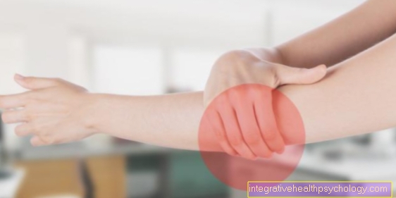
The bursa on the elbow (bursa olecrani) is there to protect the structures there (bones, tendons, ligaments and adjacent tissue). It lies in the so-called subcutaneous tissue between the skin and the bone and ensures that the skin can be moved in relation to the underlying bone. The liquid it contains compensates for shocks. It serves as a sliding layer between the structures.
Since the elbow is a highly stressed joint and can be exposed to great pressure or impact, the bursa is of great importance. If the bursa is heavily stressed, for example when the elbows are propped on the table, it can lead to inflammation (bursitis). This inflammation caused by constant stress is also known as the “student elbow”. In addition to inflammation, injuries to the bursa are not uncommon due to the exposed location.
Bursa on the hip
The bursa on the hip (bursa trochanterica) lies between the thighbone and the tendons that run over it.
There are three different types of bursa. They are located on the side of the hip between the thigh bone and three different tendons of the gluteal muscles. There are more bursa in the depth of the hip near the hip joint between the muscles there. There is also another bursa between the skin and the bone. The exact location of the hip bursa is not definitively known.
Since the hip is a highly stressed joint and can be exposed to great pressure, these bursa are important to compensate for shocks. The bursa under the tendons are important for cushioning and protecting the structures, as otherwise the tendons would lie directly on the bone. This would lead to friction damage when moving. As with the elbow, the hip bursa can be affected by inflammation.
Bursa on the knee
There are several bursae on the knee. There is a large bursa between the skin and the kneecap (bursa prepatellaris). It is flat and belongs to the skin mucous bags. It is responsible for the mobility of the skin in relation to the kneecap when the knee is bent. Since it lies very superficially, it can be injured quickly. During work that involves kneeling for a longer period of time, for example with tilers or cleaners, the permanent stress can cause the bursa to become infected.
Another large bursa (bursa suprapatellaris) lies above the knee joint. It is located between the thigh bone and the tendon of the anterior thigh muscle.He is responsible for the smooth gliding of the tendon over the bone when the knee is bent. In addition, pressures that arise in this area are evenly distributed by the bursa. There is a connection between this bursa and the joint cavity of the knee, which is why it is also known as the “suprapatellar recess”.
Another bursa (infrapatellar bursa) is located below the knee joint. It is divided into two parts. The superficial part lies between the skin and the patellar tendon. The deep part is found between the patellar tendon and the underlying bone. Like the other bursae, this two-part bursa is important for the tendon to slide smoothly over the bone. Bursa of the knee can become chronically inflamed from constant stress in a kneeling position, for example at work.
There are other smaller bursa in the hollow of the knee.
Bursa on the Achilles tendon
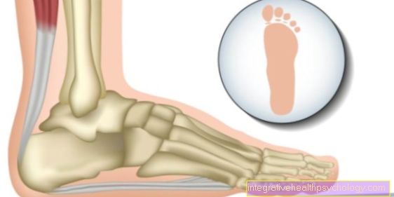
The bursa on the Achilles tendon is divided into two parts. A distinction is made between the superficial and the deep part. The superficial part is between the skin and the tendon and the deep part is between the Achilles tendon and the underlying bone. Depending on the position of the feet, the pressure on the bursa can be increased or decreased. The bursa with the fluid it contains is responsible for the even distribution of pressure and the smooth sliding of the tendon and bone.
The bursa can be chronically inflamed and extremely painful. If so, it should be surgically removed. An inflammation of the bursa can be promoted by a foot misalignment.
Bursa of the shoulder
Several bursae are found on the shoulder. A bursa lies between what is known as the ankle joint and the tendon of a shoulder muscle (bursa subacromialis). He is responsible for ensuring that the humerus does not hit the bony roof of the shoulder when the arm is raised. It is also important for the tendon to move relative to the bone.
The tendon of the muscle can change degeneratively, which can lead to inflammatory processes of the bursa. The bursa can stick together so that it can no longer fulfill its function. More bursae sit under the tendons of the shoulder muscles. Together, the bursa ensure that the muscles or their tendons slide smoothly over the bones when the arm is raised.
When working with your arms above your head, the risk of these bursa becoming infected is increased. Examples are chopping wood or painting. Inflammation can also occur during sports that involve raising the arm frequently.











.jpg)