Tooth structure
introduction

The human set of teeth contains 28 teeth in adults, 32 with wisdom teeth. The shape of the teeth varies depending on their position. Incisors are a bit narrower, but molars are all the more massive, which depends on their respective function. The structure, i.e. what the tooth is made of, is the same for every tooth and person. The hardest substance in the entire body is in our mouth, but once we have lost it, it does not come back.
The external structure
Viewed from the outside, the tooth can first be divided into three sections.
- The part that is visible and protruding from the gum is the crown of the tooth.
- The neck of the tooth is attached to them,
- which represents the transition to the tooth root, which is firmly anchored in the alveolar compartment.
The last two are overgrown by the gums. The ratio of tooth crown to tooth root is approx. 1/3 to 2/3.
The reason why the tooth consists of such a hard substance is that it is exposed to strong forces every day that we do not even notice when we chew. He has to endure a daily load of 15-30kg, in extreme cases it can even be 100kg. In order to be able to accomplish this, it is made up of various substances, which are examined in more detail in the following sections.
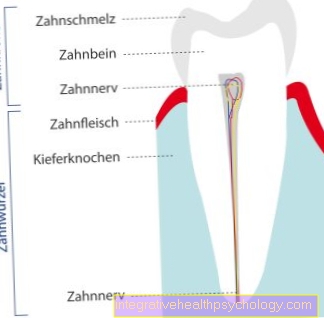
The main substance of the tooth is that Dentine, which is covered by so-called tooth enamel on the tooth neck and crown. However, the enamel is no longer to be found in the root area. There the dentin is dated Root cement enveloped. The transition from tooth enamel to root cement is at the tooth neck. The inside of the tooth consists of the pulp cavity, the supply center of the tooth.
Figure anatomy tooth

a - tooth crown - Corona dentis
b - tooth neck - Cervix dentis
c - tooth root - Radix dentis
- Tooth enamel -
Enamelum - Dentin (= dentine) -
Dentinum - Tooth pulp in the tooth cavity -
Pulp dentis in Cavitas dentis - Gums -
Gingiva - Root canal
- Cement -
Cementum - Root skin - Periodontium
- Opening of the tooth root tip -
Foramen apicale dentis - Nerve fibers
- Alveolar bone (tooth-bearing
Part of the jawbone) -
Pars alveolaris
(Alveolar process) - Blood vessels
- Tooth Root Tip -
Apex denitis - Point of division of the tooth roots
(Fork) - Bifurcation - Tooth furrow
You can find an overview of all Dr-Gumpert images at: medical illustrations
The internal structure
If you explore the tooth from the inside out, you will come across the first Tooth pulp. As mentioned above, this is the supply center for the teeth. Your tasks are the Diet, sensitivity, defense and formation. It gives the tooth its shape, nourishes it, possesses defenses and enables feeling. They can be divided into an inner and an outer zone. They are on the very outside, i.e. on the border with the dentine Odontoblastic bodythat make up the dentin. They thus line the edge of the cave from the inside. The pulp tapers towards the bottom Apical foramen. Through this the supplying vessels and nerves get into the tooth.
The next stop on the exploration tour is that Dentine. It is too 70% from minerals, such as calcium and phosphate, 20% from organic substanceswhat is mainly collagen, and 10% water. Tiny tubules can be seen in the dentine, which Dentinal tubules. The Tomes fibers lie in them. These are the processes of the odontoblasts that line the edge of the pulp cavity. The density and also the diameter of the tubules decrease with increasing distance from the pulp. The dentin, which is very close to the pulp, is called predentin because it is still uncalcified. This is followed by the circumpulpal dentin, which makes up the bulk of the dentin. Near the enamel is the third layer, the coat dentine. This has a lot of collagen fibers, is highly branched and less densely mineralized. If you were to cut the dentin crosswise, you can see certain growth lines (from Ebner lines) that are less mineralized. Depending on when the dentin is formed, one can distinguish between three types. There that would be Primary dentistthat arises during tooth development. Secondary dentist forms after the tooth root formation. Tertiary dentine always develops when the tooth has been damaged by, among other things, irritation.
The dentin in the area of the tooth crown is surrounded by tooth enamel. This exists 95% minerals, 4% water and 1% from organic substances. The enamel is formed during the development of the Ameloblasts and has a crystalline structure. The individual crystallites have a hexagonal structure and are bundled together to form several. Such bundles are called Enamel prisms. The individual enamel prisms interlock with one another. Due to the curved course of the enamel prisms, when the light is refracted, dark (diazonia) and light (parazonia) stripes are created. In enamel, growth lines are called Retzius stripes. Tooth enamel itself has no metabolism. A De- and remineralization takes place anyway, even if the ameloblasts only form enamel during development. Ions, water and dyes can pass through the enamel. The color of the enamel depends on the underlying translucent dentin. However, you can Discoloration, triggered by tea, smoke, Drugs, etc. that affect permeability.
The tooth supporting apparatus
The Teeth-supporting apparatus is also called Periodontium. Its components are that Root skin (Periodontal disease), the Root cement, the Gingiva and the Alveolar bone. The tooth-holding apparatus integrates the tooth and anchors it firmly in the bone. The root cement consists of 61% minerals, 27% organic substances and 12% water. The cement contains collagen fibers. On the one hand, these are the von Ebner fibrils and, on the other hand, the Sharpey fibers, which come from the outside of the periodontal membrane. The periodontal membrane is the last layer before the bone follows. She serves the Anchoring the tooth in the tooth socket, as well as nutrition, sensitivity and defense and consists of intertwined collagen fiber bundles. The most important cells are the Sharpey fibers, which run through the periodontal membrane. They pull from the alveolar bone to the root cement and cause the bone to be subjected to a tensile force when the teeth are loaded. The alveolar bone consists of three structures. On the one hand, the alveolar wall, which is permeable to nerves, lymph and blood vessels. The outer part is formed by the cortex and the inner part by the cancellous bone, which is filled with fat marrow.
Function of teeth
The main task that one ascribes to the teeth is theirs Chewing function. Everything we eat is chopped up by them. Almost every solid food is chewed so that it can pass through the esophagus in the next step. But that's not all. In addition to the chewing function, they also fulfill important tasks in pronunciation, i.e. the phonetics. A misaligned tooth can lead to incorrect sound formation, such as lisp. But also that To sing, Laugh and Making music would not be possible without our 28 little helpers. In addition to these tasks, they also fulfill an important role aesthetic feature. Healthy, white and vital teeth make a face appear much more attractive and personable. They are a characteristic of vitality and health. Thus, daily dental care is not only a removal of leftovers, but also a kind of beauty treatment.
Outline of the dentition
If one includes the wisdom teeth, a full-grown person has 16 teeth in the upper jaw and 16 teeth in the lower jaw. The foremost teeth are that Incisors, the Dentes incisivi decidui. They're the first two on each page. The third tooth is the canine, the Dens caninus decidui. After this tooth follow the Premolars (Dentes premolares), the 4th and 5th tooth, followed by the Molars (Dentes molares; 6, 7 and 8). The incisors are used to bite off the food, the molars to chop it up.
Summary
Even if the external appearance of the teeth does not suggest a complex structure, their individual components make them very structured and functionally appropriate so that they can cope with the daily tasks that we expect them to do. Mostly for many decades they faithfully chop our food, withstand great forces and conjure up a beautiful smile on our faces.

















.jpg)