Assessment of the liver using MRI
introduction
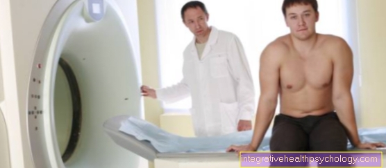
During a magnetic resonance scan (MRI), the patient is pushed into a tube that is fitted with magnetic coils. With the help of electricity, a magnetic field begins to build up, which then uses complex computing processes to generate an image.

indication
An MRI of the liver is performed whenever other imaging tests cannot provide an accurate representation of the liver. In general it can be said that the representation of Soft tissues and annoy, Tendons etc. with the MRI better can be represented as e.g. in the Computed Tomography.
A X-ray image has in the appearance of the liver no sensebecause mainly only bone can be displayed with the normal X-ray.
Especially when it is unclear Discomfort in the right upper abdomen should be at a CT or MRI can be thought of as further diagnostics. Especially when the Liver values are increased in the blood and no cause can be found, an MRI scan of the liver should be considered.
An MRI scan of the liver should also be performed whenever the Ultrasonic an unclear structure of the liver tissue was seen and could not be assigned. The MRI examination takes a long time compared to other imaging procedures. An MRI scan of the liver should take around 15-30 minutes. An MRI examination becomes difficult if the patient is under fear, especially under Claustrophobia suffers. But also a MRI for claustrophobia is possible.
An MRI examination does not have to be performed on an empty stomach per se. If, for example, an examination of the intestines or stomach is to be carried out, then it must be ensured that the patient is fasted. Otherwise this is not necessarily important. In the case of a liver examination by an MRI, the patient must not sober it can be in order to avoid corresponding air overlaps but recommended become. In this case, however, it is sufficient to not eat anything 4 hours beforehand.
MRI sober
An MRI examination does not have to be performed on an empty stomach per se. If, for example, an examination of the intestines or stomach is to be carried out, then it must be ensured that the patient is fasted. Otherwise this is not necessarily important. In the case of a liver examination by an MRI, the patient must not sober it can be in order to avoid corresponding air overlaps but recommended become. In this case, however, it is sufficient to not eat anything 4 hours beforehand.
MRI examination with or without contrast agent?
Magnetic resonance imaging can be done with or without prior Administration of contrast medium be performed. Many tissues often look similar or the same in the MRI image. In order to be able to differentiate between them better, it can be useful before the MRI examination via the vein to apply a contrast medium to the patient. This floods into the body within a few seconds and "Colors" some organs in a different signal, which then differs from the non-“stained” tissue in the resulting MRT image.
There are some recordings that are made almost exclusively with contrast medium. These include e.g. Recordings of the central nervous system and des Brain.
In the case of an MRI of the liver, it can be considered whether the application of contrast medium is necessary. It should be noted namely that contrast media is also not infrequently allergic reactions can lead. For this reason, it is very important to inquire about a known allergy before the MRI examination. In case of a MRI examination The liver can also first take one or more images without contrast agent and see whether the view is sufficient. If this is not the case, contrast media can also be applied later.
Contrast medium administration by Primovist
Primovist is a contrast medium that is mainly used for MRI examinations of the liver. It consists of Gadolinium and is mainly used in the diagnosis of Liver metastases or des Liver cancer used.
When using Primovist, it is important that not only the actual abnormalities in the liver are better represented because the corresponding tissue is "stained" and separated from the rest of the tissue, but it also allows statements to be made as to whether it is benign or benign malignant mass.
Duration of an MRI examination
The duration varies greatly depending on the organ section to be examined. In principle, however, one can say that an MRI examination usually takes longer than a CT examination or an X-ray. For example, if a Spine on MRI If the device is to be viewed more closely, the patient must already have a duration of up to 30 minutes calculate.
The radiologist monitors the image quality during the exposure. If there are inaccuracies or blurring in the image section, certain recording sequences can be repeated accordingly, which leads to an extension of the overall diagnosis. A whole It is therefore difficult to give an exact time.
The very common ones MRI examinations of the shoulder e.g. usually do not take that long while the representation of the abdominal area can take up to 30 minutes.
If a native image is first taken (i.e. the examination is carried out without contrast agent) and then contrast agent is injected into the vein, the total time of the MRI examination is also delayed.
During the MRI examination of the liver should, if all sections are to be displayed with and without contrast agent, also with a length of approx. 15 to 30 minutes calculate. If the patient is claustrophobic, a low dose should be considered Sedatives (e.g. Tavor to give).
MRI costs

An MRT examination is one of the more expensive examination instruments in medicine. In addition to very high development and acquisition costs, this is also due to the high expenditure on maintenance and repair.
In contrast to an X-ray or CT examination, an MRT examination is harmless, but costs, length and necessity should be taken into account when establishing the indication.
The cost of an MRI of the head and neck region is around EUR 370. If the administration of contrast agent is also necessary, the costs increase to 445 EUR.
Examination of the spine with contrast medium would cost EUR 430 and the examination of the chest organs would cost EUR 360 without contrast medium and EUR 440 with contrast medium.
The examination of the shoulder region and the MRI of the cervical spine are the most expensive examinations.Without contrast agent, costs of 517 EUR are due with contrast agent costs of almost 600 EUR.
As a rule, the costs are covered by the statutory health insurance. In cases of doubt, an application for reimbursement must be submitted to the statutory health insurance before an examination.
Read more about this topic under Costs of an MRI examination.
MRI of the gallbladder
An MRI examination of the bile is always carried out if abnormalities have been seen in the ultrasound that cannot be reliably assigned. Even if Gallstones in the Gallbladder and especially in Bile duct were seen, an MRI scan should show the exact location of the gallstone.
Increase in bile values in the Blood count and inconspicuous ultrasound can just as easily necessitate an MRI scan of the gallbladder as it does a Space claimfound in the area of the gallbladder wall or in the gallbladder.
Quite often, an MRI scan of the gallbladder is also ordered if a Occlusion of the gallbladder or bile duct it is feared but the cause is not clear. Malignant neoplasms can then be seen very well with the help of the MRI examination.
It is important to note that there may be delays and long waiting times can come when a general decision is made to have an MRI examination. Sometimes up to 4 weeks can pass before an appointment for an MRI scan is available. It is important for the family doctor to decide whether there is enough time or a faster appointment needs to be found.
Will an MRI scan of the Gallbladder and or the bile ducts should be carried out with a 10-20 minute length be expected from the investigation. Often, native recordings are first made without contrast agent and then the recordings are repeated with an amount of contrast agent released into the veins.
Hemangioma
At a Hemangioma it is a vascular remodeling. This can occur in all areas where Blood vessels lie. Hemangiomas are very common in the face area. But they can also occur in the liver. As a rule, the person concerned does not know anything about them because they no symptoms do.
The first clue is usually a random ultrasound examination of the abdomen, in which circular, whitish areas are seen in the liver area. These patchy changes are usually already through a Eye diagnosis to be classified as a hemangioma. If you are not sure, an MRI scan of the liver can also be performed.
A hemangioma is important to be distinguished from liver metastasis. A hemangioma is harmless and needed usually no treatment. It is also important to observe the size and number of hemangiomas. A Ultrasound check-up once a year by the family doctor is usually sufficient. Hemangiomas in visible areas can be surgically removed for cosmetic reasons. In the area of the internal organs, such as the liver, this makes no sense. If the hemangiomas are on the liver surface, they can in principle tear open and bleed due to mechanical shear forces. However, this is so rare that a prophylactic removal tends to make no sense power.
MRI procedure
The patient for whom an MRI scan is to be performed walks with his Transfer from the family doctor with the MRI liver on it to a radiologist with whom an appointment has been made beforehand. Often a waiting period It may take up to 4 weeks before an MRI scan can be performed.
In advance, the patient must have a Information sheet read through and sign. The MRI examination is one safe investigation, however, it may be necessary Contrast media to inject what in principle to allergic reactions can lead. It is also important to inform the radiologist whether there are metallic implants in the body. In Germany an MRT examination is allowed Pacemaker wearers do not as there is a risk of electrode loosening.
The patient then has to lie down on a couch that is automatically inserted into the MRI machine. Before that he gets another one venous access placed in the arm vein in case contrast agent has to be injected. As it goes through the electrical coils extremely loud in an MRI machine, the patient gets one headphone put on your ears. Using these headphones, he can also receive instructions from the control room where a medical technical assistant or radiologist will carry out the examination.
Through a Button in hand can the patient end the examination in an emergencyif he's not okay. The examination can take up to 30 minutes.
Read more about this under Procedure of an MRI.

