Conjunctiva
synonym
Medical: Sclera conjunctiva
lat .: Conjunctiva
definition
The conjunctiva is a part of the eye. As a mucous membrane, it rests on part of the outside of the eyeball and on the inside of the eyelids. It can be changed in the context of diseases, this can be recognized primarily by its color.

anatomy
The conjunctiva (Conjunctiva) consists of two parts.
- The conjunctiva tarsi (the Tarsus is part of the eyelids) covers the inside of the upper and lower eyelids as the outermost layer.
- The conjunctiva bulbi (the B.ulbus oculi is the eyeball) covers from the outside the part of the eyeball that is not covered by the cornea, i.e. the upper and lower edge, where the dermis (Sclera) runs.
A multilayered, non-keratinizing squamous epithelium with mucus-producing goblet cells forms the basic structure of the conjunctiva. The change between keratinizing squamous epithelium of the skin (epidermis) in the non-keratinizing squamous epithelium of the conjunctiva lies on the conjunctiva tarsi.
At the fornix superior (upper bulge), which is located in the depth of the eye socket, the conjunctiva tarsi overlaps from the eyelid into the conjunctiva bulbi of the eyeball. The same applies to the inferior fornix, the lower bulge. The conjunctival sac is formed in these areas.
The conjunctiva is transparent and very well supplied with blood. It is firmly attached to the eyelids, while it is only loosely attached to the eyeball. The conjunctiva is sensitively innervated by small nerve fibers, which are all branches of the trigeminal nerve (5th cranial nerve):
- Frontal nerve
- Lacrimal nerve
- Infraorbital nerve and
- Nasociliary nerve
The arterial vascular supply takes place via branches of the Ophthalmic artery.
Special structures of the conjunctiva of the eye:
- The so-called plica semilunaris is a mucosal duplication that is tender, soft and flexible at the inner corner of the eye.
- A caruncle is a tissue protrusion between the plica semilunaris and the inner corner of the eyelid, it consists of mucous membrane, skin parts and sebum glands.
- The goblet cells that secrete mucus are present throughout the conjunctival epithelium.
- Acessory tear glands supply the aqueous component of the tear film and are located on the upper edge of the so-called tarsal plate of the upper eyelid and in the area of the fornices.
What is the conjunctival sac
The conjunctival sac is also known as the conjunctival sac and is an anatomical structure in every person that is located both between the inside of the upper eyelid and the eyeball and between the inside of the lower eyelid and the eyeball. Therefore one can differentiate between an upper and lower conjunctival sac.
The conjunctival sac is formed by the folds of the envelope of the various parts of the conjunctiva and the adjacent cornea and is also used in anatomy Fornix conjunctivae called. This is where the conjunctiva, which covers the back surface of the eyelids, turns over and then forms the conjunctiva that covers the eyeball.
In healthy people there is always a certain amount of tear fluid in the conjunctival sac, which keeps the surface moist and supple and protects against infections. Medicines can also be applied here in ophthalmology. If the eye is diseased, pus or foreign bodies can be found here, for example, which disrupt the normal function of the conjunctiva and the eye.
Read more on the subject at: Conjunctival sac

- Cornea - Cornea
- Dermis - Sclera
- Iris - iris
- Radiant Bodies - Corpus ciliary
- Choroid - Choroid
- Retina - retina
- Anterior chamber of the eye -
Camera anterior - Chamber angle -
Angulus irodocomealis - Posterior chamber of the eye -
Camera posterior - Eye lens - Lens
- Vitreous - Corpus vitreum
- Yellow spot - Macula lutea
- Blind spot -
Discus nervi optici - Optic nerve (2nd cranial nerve) -
Optic nerve - Main line of sight - Axis opticus
- Axis of the eyeball - Axis bulbi
- Lateral rectus eye muscle -
Lateral rectus muscle - Inner rectus eye muscle -
Medial rectus muscle
You can find an overview of all Dr-Gumpert images at: medical illustrations
histology
The conjunctiva consists of a multi-layered, highly prismatic columnar epithelium in which goblet cells are embedded. The secretion of the goblet cells is part of the tear film.
Function of the conjunctiva
The conjunctiva acts as a kind of outer protective covering of the eye and contributes to the production of the tear film through the secretion of its goblet cells.
This one is for that eye vital.
Clinical fact of the conjunctiva
A closer look reveals a lot about the color of the conjunctiva. Redness may indicate conjunctivitis (Inflammation of the conjunctiva) be. A yellowish colored conjunctiva is often the first indication of jaundice (jaundice). This is caused by increased deposition of blood breakdown products. These are no longer red in color like the blood itself, but have a yellow intrinsic color.
Also an anemia (anemia) can be recognized by the conjunctiva on closer inspection. This is then paler, i.e. whiter than usual.
Conjunctivitis is also of clinical importance (Conjunctivitis). It can arise in the context of local processes (e.g. foreign bodies in the eye) but also in systemic reactions (e.g. bacterial infection). Allergic rhinoconjunctivitis, better known as hay fever, is also very common.
Conjunctival diseases
Acute bacterial conjunctivitis
Conjunctivitis (Conjunctivitis) can in principle be triggered by numerous pathogens, but only a few are able to cause severe acute conjunctivitis in healthy people (streptococci, Corynebacterium diphteriae, Neisserien, Haemophilus).
Staphylococcus aureaus, Streptococcus pneumoniae and Haemophilus aegypticus are the most common causative agents of catarrhal conjunctivitis. Infection can occur in a number of ways: air, gastrointestinal tract, and many more.
Typical of an infection with Haemophilus influenzae and Corynebacterium diptheriae is a pronounced swelling of the eyelids. On the other hand, membranes are mainly formed with infections Streptococcus pyogenes and Corynebacterium diphtheriae. The so-called petechiale (punctiform) bleeding in the eyelids are with infections Streptococcus pneumoniae and H. influenzae.
If the conjunctiva is inflamed, there is usually no swelling of the lymph nodes or skin involvement. Complications are severe keratitis (inflammation of the cornea) (especially with Corynebacterium diphtheriae, Neisseries, H. aegypticus), Sepsis (Corynebacterium diphtheriae, Neisseries, Haemophilus, Pseudomonas) Dacryocystitis and scarring.
The choice of the appropriate therapy depends on the severity: A slight conjunctivitis (conjunctivitis) is usually treated with local antibiotics (gentamycin, erythromycin, chloramphenicol, neomycin, gatifloxacin, levofloxacin, ofloxacin, ciprofloxacin, etc.) without a smear and determination of the exact pathogen treated with eye drops or antibiotic ointments.
also read: Floxal eye ointment
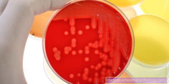
In severe conjunctivitis associated with eyelid swelling, massive secretion, membrane formation and possibly also corneal inflammation (keratitis), the pathogen is determined by smear, Gram and Giemsa staining and culture of the pathogen on blood and so-called chocolate agar. At the beginning, if the exact pathogen has not yet been determined, treatment is carried out with highly concentrated antibiotics (gentamycin, ceftazidime 5%) and then later the treatment is adapted to the exact resistances of the pathogen present. If necessary, irrigation or cycloplegia (paralysis of the ciliary muscle, which leads to paralysis of the accommodation of the eye and mydriasis; e.g. medicinally) of the eye is performed.
Symptoms of conjunctivitis:
Classic signs that indicate conjunctivitis are:
- Burn
- itch
- light pain
- white or yellow secretion
- Redness
- Photosensitivity
- swelling
- Papillae (the ophthalmologist sees with the help of the slit lamp)
- glued eyelids
Gonococcal conjunctivitis
The causative agent of this conjunctivitis is the aerobic, gram-negative diplococcus (N. gonorrhoeae), with preference for the mucous membrane and genital tract. The culture is ideally carried out with slightly increased CO2 pressure on so-called chocolate agar or Thayer-Martin medium. It is important between N. gonorrhoeae and N. meningitidis to distinguish.
In adults, infection usually occurs through self-contamination. Gonococcal conjunctivitis can lead to severe keratitis (inflammation of the cornea), possibly with perforation, to sepsis, arthritis and dacroadenitis (inflammation of the lacrimal gland).
In addition to various prophylactic agents, a culture is created to treat the disease itself. Inpatient treatment and isolation of those affected makes sense. Frequent rinsing of the affected eye with isotonic saline solution facilitates healing. In addition, the antibiotic erythromycin is given for topical application and the antibiotic ceftriaxone, penicillin or spectinomycin is given parenterally (as an infusion) for 7-14 days. The sexual partner must also be treated with gonococcal disease in order to prevent a possible ping-pong effect. If the diagnosis is uncertain, chlamydia must also be treated.
What is a conjunctival cyst?
A conjunctival cyst is a harmless disease of the eye that is relatively common and usually does not cause any problems. This is a bulging of the conjunctival surface. These often occur after inflammation or injury. As a rule, serous, i.e. clear and not viscous, fluid of varying proportions accumulates under the bulge.
The conjunctival cyst is usually so small that it does not cause any problems. In some cases, however, it happens that the movement of the eyeball feels strange or difficult and one has a proven foreign body sensation. In this case, an ophthalmological check should definitely be carried out. If in doubt, this should generally be done.
After the examination by the ophthalmologist, the conjunctival cyst is usually punctured. This means that it is pierced and emptied with the help of a needle. This is usually done under a local anesthetic and should never be done by yourself. This is not a painful process. Complications are extremely rare. However, if inflammation occurs as a result, the doctor should definitely be consulted again.
However, after removal of the conjunctival cyst, recurrences often occur. This means that the conjunctival cyst recurs relatively often and may cause problems again. In this case, the doctor can be consulted again.
Read more on the subject at: Conjunctival cyst
What is conjunctival irritation?
There are many different causes of conjunctival irritation, all of which can cause similar symptoms. Conjunctival irritation should not be equated with conjunctivitis. However, conjunctivitis can irritate the conjunctiva and cause the same symptoms.
In the context of conjunctival irritation, an inflammatory reaction occurs, which results in increased blood flow. Thus, irritation of the conjunctiva typically leads to reddening of the eye, which is accompanied by increased tear secretion. In contrast to irritation of the cornea, irritation of the conjunctiva is not painful. There is also no decrease in visual acuity. Additional symptoms can occur, but they do not occur in all cases. For example, a foreign body sensation or purulent secretion should be mentioned here.
Possible causes of conjunctival irritation are a superficial injury, slight infection, an allergy or other systemic diseases. In this case, if it occurs more frequently or for a long time, a doctor should be consulted for clarification.
Swollen conjunctiva
A swollen conjunctiva is used in medical terminology as well Chemosis called. In the case of chemosis, as part of pathological processes, fluid accumulates in and under the conjunctiva, known as edema, which makes it appear swollen and stands out from the layers below. The conjunctival edema can lead to either a milky-white opacity or severe reddening of the conjunctiva. In addition, there is pain and possibly a decrease in visual acuity.
In addition to inflammation caused by bacteria or viruses, the cause of a swollen conjunctiva can also be irritation of the conjunctiva. This can happen through superficial damage such as foreign bodies, trauma or UV radiation as well as allergies. Wearing contact lenses for too long can also be the cause. If there is a problem with the outflow of blood or the lymph in the eye socket, the increased pressure can also lead to the development of conjunctival edema. This drainage disorder occurs, for example, after a trauma or with a tumor. However, these reasons are rare.
Therapy by the doctor takes place depending on the cause. If inflammation is the reason, it is treated. In the case of allergies, attempts are made to avoid the trigger. Superficial damage to the conjunctiva can be treated with rest, soft contact lenses or, in severe cases, surgery.
Conjunctival tumor
Conjunctival tumors are a rare condition that affects the conjunctiva of the eye. In contrast to other tumors, however, a conjunctival tumor is usually benign and therefore easy to remove and treat, which means that, as a rule, no long-term damage and negative effects occur. Nevertheless, malignant, i.e. malignant, tumors occur now and then.
Even one conjunctival cyst can be counted as a conjunctival tumor. The formation of new blood vessels in the conjunctiva, a so-called hemangioma, is also known as a tumor. Although it doesn't look nice, it hardly causes any discomfort and is easy to treat. In children, this tumor can even go away on its own. In adults, a hemangioma is surgically removed.
Other benign conjunctival tumors are melanosis and the conjunctival nevus. However, both show a certain risk of degeneration, so that they must be checked regularly to prevent damage at an early stage. A conjunctival nevus corresponds to a birthmark that is located on the eye. Melanosis is caused by too much build-up of dark skin pigment.
Malignant conjunctival tumors are carcinoma and lymphoma. Carcinoma is caused by degenerate epithelial cells, while lymphoma arises from cells of the immune system. These do not always express themselves in the same way (changed surface, pain, foreign body sensation) and are sometimes recognized too late. Therapy consists of surgical removal for carcinoma and radiation therapy for both tumors.
Read more on the subject at: Conjunctival tumors
Conjunctival melanoma
The conjunctival melanoma represents the malignant degeneration of the melanosis or a conjunctival nevus. Also due to the frequent controls of the conjunctival nevus or the melanosis, the conjunctival melanoma is a rare, but nevertheless serious disease and requires early and decisive therapy.
The conjunctival melanoma is noticeable by a dark spot in the area of the conjunctiva, which is usually thickened and protruding. The area around the conjunctival melanoma is often darkened and has a high density of blood vessels.
The doctor makes the diagnosis based on a clinical examination and tissue analysis through histology. In order to rule out a spread to the nearby lymph nodes, a CT or MRI is made. A metastasis in the rest of the body should also be ruled out if there is justified suspicion.
Treatment consists of surgical removal followed by radio or chemotherapy. Since the tumor often recurs, close follow-up care is recommended.
Read more on the subject at: Conjunctival tumor
Conjunctival lymphoma
Conjunctival lymphoma is a rare tumor found in the human eye. In contrast to most other tumors, conjunctival lymphoma is malignant and requires therapy. However, the prognosis is good.
The conjunctival lymphoma is noticeable through a painless swelling in the area of the conjunctiva. This is usually slightly reddish and localized on the conjunctiva of the lower eyelid. It arises from degenerate cells of the immune system and can therefore arise both locally and elsewhere in the body.
Therapy should start as early as possible. Due to the different causes and the different places of origin, the therapy can vary greatly. Radiotherapy, chemotherapy and therapy with so-called biologicals can be considered for this.
Conjunctival hemorrhage
Conjunctival hemorrhage is a relatively common clinical picture, but it is usually harmless. It can arise from many possible causes and usually does not cause any problems.
Conjunctival hemorrhage is noticeable as a visible, red spot on the conjunctiva. The bleeding is not painful and does not cause visual disturbances. Only a slight irritation of the conjunctiva sometimes occurs. Often it occurs when the pressure inside the eye or blood vessels increases. This is the case with coughing, sneezing, straining, vomiting, exercising, but also with childbirth and high blood pressure. Rubbing the eyes too hard can also cause bleeding.
With drug anticoagulation, conjunctival bleeding can also occur more frequently. This then largely affects the elderly. Contact lenses or an injury can also be a possible cause.
Conjunctival hemorrhage resolves on its own within a few days to two weeks and does not require any therapy. Therapy should only be considered for underlying systemic diseases such as high blood pressure or some metabolic diseases such as diabetes mellitus.
Conjunctival tear
A conjunctival tear is a relatively common disease that usually does not have any serious consequences. An external mechanical load is the first to injure the conjunctiva. This manifests itself as a feeling of foreign body, slight pain and bleeding. There may also be an increased secretion of tear fluid.
While small tears in the conjunctiva heal on their own, large tears are treated by sewing the edges of the wound together. If the affected area becomes inflamed, a doctor should definitely be consulted.



.jpg)
